
Fig. 3
- ID
- ZDB-FIG-240307-13
- Publication
- Russell et al., 2021 - Pathogenic effect of TP73 Gene Variants in People With Amyotrophic Lateral Sclerosis
- Other Figures
- All Figure Page
- Back to All Figure Page
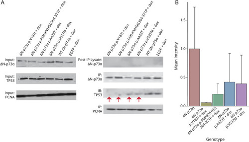
ΔN-p73α Containing ALS Patient Mutations Expressed in Neuro-2A Cells Displayed Impeded Binding to Proapoptotic p53 (A) Western blots show equal input level of protein (proliferating cell nuclear antigen [PCNA], ΔN-p73α, and p53) before immunoprecipitation. Protein was immunoprecipitated with ΔN-p73α or PCNA antibody. Postimmunoprecipitation lysate demonstrates that all ΔN-p73α protein was immunoprecipitated with ΔN-p73α antibody. Blots were probed for ΔN-p73α, p53, and PCNA. PCNA was used as a loading control and was immunoprecipitated in tandem to ΔN-p73α. Red arrows denote less intense p53 protein bands immunoprecipitated by mutant ΔN-p73α compared to wild-type (WT) ΔN-p73α and enhanced green fluorescent protein (EGFP) (in EGFP lane, ΔN-p73α band is endogenous protein). (B) Quantification of p53 band intensity of Western blots performed after ΔN-p73α immunoprecipitation. Shown are mean intensity and SD for p53 bands from coimmunoprecipitations performed in biological duplicate for Neuro-2A cells expressing ST ΔN-p73α or mutant ΔN-p73α on doxycycline (dox) exposure. ALS = amyotrophic lateral sclerosis. |