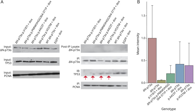
Fig. 3 ΔN-p73α Containing ALS Patient Mutations Expressed in Neuro-2A Cells Displayed Impeded Binding to Proapoptotic p53 (A) Western blots show equal input level of protein (proliferating cell nuclear antigen [PCNA], ΔN-p73α, and p53) before immunoprecipitation. Protein was immunoprecipitated with ΔN-p73α or PCNA antibody. Postimmunoprecipitation lysate demonstrates that all ΔN-p73α protein was immunoprecipitated with ΔN-p73α antibody. Blots were probed for ΔN-p73α, p53, and PCNA. PCNA was used as a loading control and was immunoprecipitated in tandem to ΔN-p73α. Red arrows denote less intense p53 protein bands immunoprecipitated by mutant ΔN-p73α compared to wild-type (WT) ΔN-p73α and enhanced green fluorescent protein (EGFP) (in EGFP lane, ΔN-p73α band is endogenous protein). (B) Quantification of p53 band intensity of Western blots performed after ΔN-p73α immunoprecipitation. Shown are mean intensity and SD for p53 bands from coimmunoprecipitations performed in biological duplicate for Neuro-2A cells expressing ST ΔN-p73α or mutant ΔN-p73α on doxycycline (dox) exposure. ALS = amyotrophic lateral sclerosis.
Image
Figure Caption
Acknowledgments
This image is the copyrighted work of the attributed author or publisher, and
ZFIN has permission only to display this image to its users.
Additional permissions should be obtained from the applicable author or publisher of the image.
Full text @ Neurology

