FIGURE

Fig 1

ID
ZDB-FIG-240306-43
Publication
Li et al., 2024 - Defining morphologically and genetically distinct GAGBergic/cholinergic amacrine cell subtypes in the vertebrate retina
Other Figures
All Figure Page
Back to All Figure Page
Fig 1

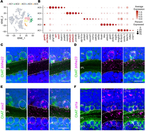
Single-cell RNA-seq identifies 2 GABAergic/cholinergic AC types.

(A) t-SNE plot showing 5 clusters of ACs (in colors) in single-cell RNA-seq data. (B) Expression pattern of marker genes for each cluster in (A). Marker genes gad1b (GABAergic), vachta (cholinergic), sox2 (AC5), bhlhe22 (AC4), glyt1 (AC3), gad2 (AC2), and sox4a (AC1) are highlighted in red. (C-F) Validation of marker TFs for cluster AC4 (C and D) and AC5 (E and F) using in situ hybridization (magenta) combined with ChAT immunostaining (green) of the 5-dpf zebrafish. Dashed circles, ACs with positive signals. Scale bars, 10 μm. AC, amacrine cell; ChAT, choline acetyltransferase; dpf, days post-fertilization; GCL, ganglion cell layer; INL, inner nuclear layer; IPL, inner plexiform layer; TF, transcription factor.

Expression Data

Expression Detail
Antibody Labeling
Phenotype Data

Phenotype Detail
Acknowledgments
This image is the copyrighted work of the attributed author or publisher, and ZFIN has permission only to display this image to its users. Additional permissions should be obtained from the applicable author or publisher of the image. Full text @ PLoS Biol.