|
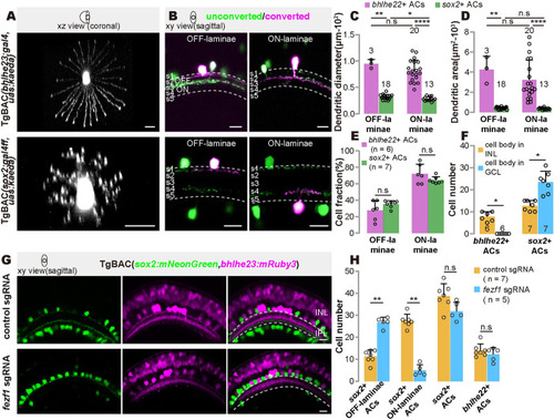
Morphological characteristics of 2 GABAergic/cholinergic AC types. (A) Representative images showing the dendrites of bhlhe22+ type and sox2+ type. (B) Representative images showing the cell body positioning and dendritic arbors of ON- and OFF-laminae of bhlhe22+ type and sox2+ type. (C and D) Quantification of the dendrite diameters (C) and sizes (D) in (A). Each circle represents one cell at 5 dpf. (E) Quantification of cell fraction of ON- to OFF-laminae in (B). (F) Quantification of the number of cells with cell body located in INL and GCL of bhlhe22+ and sox2+ type. (G) Representative images of bhlhe22+ and sox2+ type after fezf1 disruption. (H) Quantification of the number of ON- and OFF-laminae subtype of sox2+ cells, total sox2+ cells, and total bhlhe22+ cells in (G). Each circle represents one fish at 5 dpf. The data underlying this figure can be found in S3 Data. Scale bars, 10 μm. AC, amacrine cell; dpf, days post-fertilization; GCL, ganglion cell layer; INL, inner nuclear layer; sgRNA, small guide RNA.
|