
Fig. 4
- ID
- ZDB-FIG-240131-22
- Publication
- Cao et al., 2023 - Late fetal hematopoietic failure results from ZBTB11 deficiency despite abundant HSC specification
- Other Figures
- All Figure Page
- Back to All Figure Page
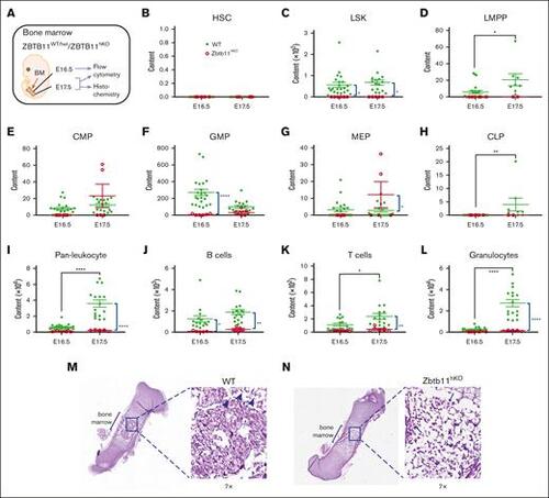
The absence of Zbtb11 in the hematopoietic compartment results in fetal BM failure. (A) Schematic description of the analysis. Scatter plots showing absolute quantification of total BM content at E16.5 and E17.5: (B) HSC, (C) LSK, (D) LMPP, (E) CMP, (F) GMP, (G) MEP (H), CLP, (I) pan leukocytes, (J) B cells, (K) T cells, and (L) granulocytes at E16.5 and E17.5. For panels B, C, E, F, G, I, and L: E16.5 WT, n = 22 and KO, n = 6; E17.5 WT, n = 16 description of the KO, n = 5. For panels D and H: E16.5 WT, n = 22 and KO, n = 6; E17.5 WT, n = 8 and KO, n = 2; For panels J and K: E16.5 WT, n = 15 and KO, n = 5; E17.5 WT, n = 16 and KO, n = 5. Hematoxylin and eosin staining of E17.5 femur sections for (M) WT and (N) Zbtb11hKO. Insets are original magnification ×7 of boxed area. Arrowheads indicate enucleated erythroid cells (M), and arrows indicate nucleated erythroid cells (N). WT, wild-type controls (green); KO, Zbtb11hKO (red); E, embryonic day; Data ± standard error of the mean. Two-way analysis of variance with Šídák multiple comparisons: ∗P < .05; ∗∗P < .01; ∗∗∗∗P < .0001. |