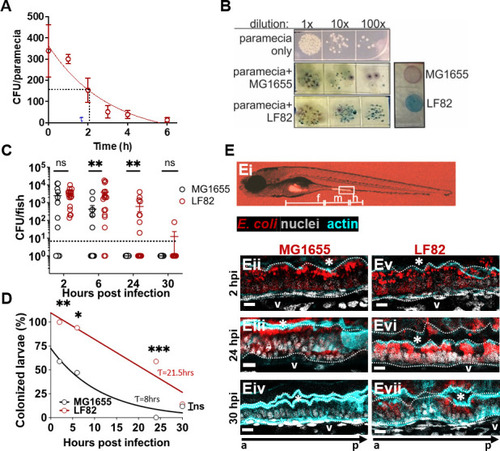
AIEC LF82 colonizes the larval zebrafish intestine better than MG1655. (A) AIEC-loaded paramecia sampled from 0 to 6 hours post incubation, and CFU/paramecia was calculated. AIEC half-life (τ) in paramecia is 2.1 hours. Data are means ± SEM, n = 3. (B) Bacterial colonies from tissue homogenates grown on CHROMagar O157. The zebrafish microbiota (white colonies) can be distinguished from AIEC LF82 (dark blue colonies) and E. coli MG1655 (mauve colonies). (C) Quantification of LF82 and MG1655 CFUs/fish. Fish with CFU below the detection limit (10 CFU/fish, dashed line) were annotated as 1 CFU. Data are from individual fish (n = 14) and means ± SEM. (D) Colonized larvae (%) are the percentage of fish with a burden above the detection limit; n = 14. Non-linear regression, first-order decay, ROUT outlier test with Q = 0.2%, paired t-test and Wilcoxon test. *, P ≤ 0.05; **, P ≤ 0.01; ***, P ≤ 0.001, ns, not significant. (E) Images of larvae colonized with E. coli (red), (Ei) whole larva at 10× magnification with intestinal segments (foregut (F), midgut (M), hindgut (H)) marked. (Eii–vii) Sagittal views of the midgut of larvae colonized with MG1655 (Eii–iv) and LF82 (Ev–vii) at 2, 24, and 30 hpi. The dotted white line outlines the intestinal epithelium and separates it from the lumen, indicated by *, and the blood vessel below the basement membrane (V). a to p marks anterior to posterior orientation; Scale bars = 100 um, E. coli (red), phalloidin (cyan, cell outline), nuclei (4′,6-diamidino-2-phenylindole, white), images are representative of n = 3.
|
