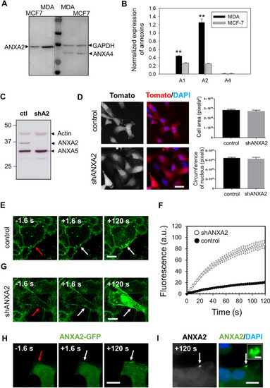
Highly expressed in MDA-MB-231 cells, ANXA2 promotes membrane repair. A Representative image of western-blot analysis showing the revelation of ANXA2 in MDA-MB-231 and MCF7 cells, as compared to GAPDH (loading control). B The histogram presents mean values (± SEM) of the ratio ANX/GAPDH from five independent experiments, analyzed by the gel analysis plugging of ImageJ. A representative membrane of the detection of ANXA1 is presented in supplementary Fig. S1. Student t test for independent samples. **p < 0.01. C ANXA2-deficient MDA-MB-231 cells were generated by shRNA transduction strategy. The cellular content of ANXA2 in MDA-MB-231 cells transduced with lentiviral particles containing shRNA targeting ANXA2 (shA2) or a scrambled shRNA (ctl) was quantified by Western blotting. D Control and shANXA2 MDA-MB-231 cells, which expressed constitutively the tdTomato fluorescent protein were imaged by fluorescence microscopy. Right-hand histograms display mean cell area (in pixels2) and nuclei circumference (in pixels) measured by the imageJ software using Tomato and DAPI images. The mean values (+ / − SEM) were calculated from at least 30 cells from three independent experiments. No statistical difference (student t test) was observed for the two parameters. Scale bar: 20 µm. E, G Sequences of representative images showing the response of a control (E) or shANXA2 (G) MDA-MB-231 cell to a membrane damage performed by 110-mW infrared laser irradiation, in the presence of FM1-43 (green). In all figures, the area of membrane irradiation is marked with a red arrow before irradiation and a white arrow after irradiation. Scale bars: 10 μm. F Kinetic data represent the FM1 − 43 fluorescence intensity for control (black filled circles) or shANXA2 (empty circles) MDA-MB-231 cells, integrated over whole cell sections, averaged for about 30 cells (+ / − SEM). H Recruitment of ANXA2 to the site of membrane injury. MDA-MB-231 cells transfected with the plasmid pA2-GFP were damaged by laser ablation. Red arrow, area before irradiation; white arrow, area after irradiation. I Subcellular localization of endogenous ANXA2 in damaged MDA-MB-231. MDA-MB-231 cells were irradiated with a 110-mW infrared laser (white arrow) in DPBS + Ca2+, then fixed and immunostained for ANXA2 and counterstained with DAPI (blue). After laser injury, MDA-MB-231 cells exhibited an accumulation of ANXA2 at the disruption site. The inset displays a magnified image of the disruption site where concentrates ANXA2. Scale bars: 10 µm on the images and 1 µm within the inset
|
