
Fig. 8
- ID
- ZDB-FIG-231212-17
- Publication
- Argitekin et al., 2023 - Dopamine-Conjugated Bovine Serum Albumin Nanoparticles Containing pH-Responsive Catechol-V(III) Coordination for In Vitro and In Vivo Drug Delivery
- Other Figures
- All Figure Page
- Back to All Figure Page
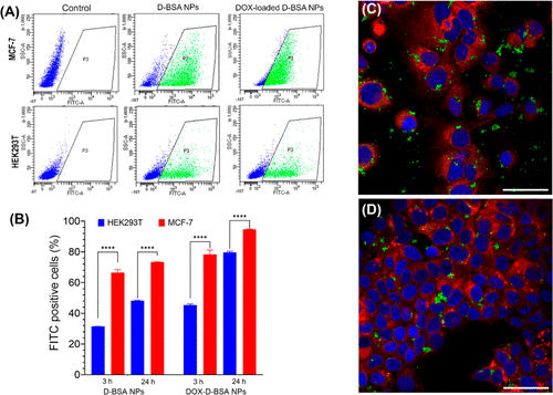
(A) Flow cytometry analysis of cellular uptake assay of FITC-labeled unloaded and loaded NPs. Both MCF-7 and HEK293T cells were treated with NPs for 3 h. Flow cytometry plots show FITC positive cells (green, region P3) and FITC negative cells (blue). (B) Ratio of cells that internalized FITC-labeled NPs were quantified from flow cytometry analysis of 100,000 events and average of two independent cell sets were plotted. NPs were internalized by MCF-7 cells more efficiently compared to HEK293T cells (****p < 0.0001). (C,D) Confocal microscopy images (40XW objective) of MCF-7 cells incubated for 24 h with 0.075 mg/mL of (C) FITC-labeled D-BSA NPs (green) or (D) DOX-loaded FITC-labeled D-BSA NPs (green). Cell membranes were stained with Dil (red) live cell dye and nuclei were stained with DAPI (blue). Scale bar = 50 μm. |