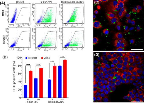
Fig. 8 (A) Flow cytometry analysis of cellular uptake assay of FITC-labeled unloaded and loaded NPs. Both MCF-7 and HEK293T cells were treated with NPs for 3 h. Flow cytometry plots show FITC positive cells (green, region P3) and FITC negative cells (blue). (B) Ratio of cells that internalized FITC-labeled NPs were quantified from flow cytometry analysis of 100,000 events and average of two independent cell sets were plotted. NPs were internalized by MCF-7 cells more efficiently compared to HEK293T cells (****p < 0.0001). (C,D) Confocal microscopy images (40XW objective) of MCF-7 cells incubated for 24 h with 0.075 mg/mL of (C) FITC-labeled D-BSA NPs (green) or (D) DOX-loaded FITC-labeled D-BSA NPs (green). Cell membranes were stained with Dil (red) live cell dye and nuclei were stained with DAPI (blue). Scale bar = 50 μm.
Image
Figure Caption
Acknowledgments
This image is the copyrighted work of the attributed author or publisher, and
ZFIN has permission only to display this image to its users.
Additional permissions should be obtained from the applicable author or publisher of the image.
Full text @ Biomacromolecules

