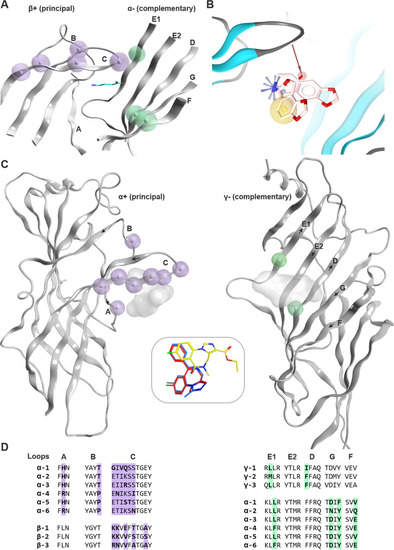
Known ECD interface binding sites for GABA and benzodiazepines: A GABA sites: β3 + / α1 − of 6HUJ showing in lilac residue positions that are different in the beta subunits (β1, β2, β3), as well as those that differ in the alpha subunits in light green. The GABA molecule is rendered in cyan sticks. More details are provided in Supplementary Figure S3. B Chemical feature interactions of the bicuculline bound 6HUK structure rendered with LigandScout 4.4 Expert. Yellow spheres, blue stars, and red vectors represent hydrophobic, positive ionizable, and hydrogen bond acceptor interactions, respectively. C Side view of the benzodiazepine binding site (α + /γ − interface) from a PDBeFold superposition of selected atomic resolution structures (PDB IDs: 6HUP—diazepam, 6HUO—alprazolam, 6D6T/6D6U—flumazenil). The subunits are rendered individually for more clarity, and the variable positions are highlighted as in panel A with lilac for the principal and light green for the complementary face, respectively. The insert box in the middle depicts the binding modes of diazepam (red), alprazolam (blue), and flumazenil (yellow). The corresponding ligands are displayed on the protein as shadows for orientation. The direction of the beta strands on the complementary face is indicated by arrows. D Partial alignment for the binding site forming segments matching panels A and B. More details on variable positions, including those that are found on segment F, are provided in Supplementary Figures S4 and S5)
|

