FIGURE SUMMARY
Title

GABAA receptor-mediated seizure liabilities: a mixed-methods screening approach

Authors
Bampali, K., Koniuszewski, F., Vogel, F.D., Fabjan, J., Andronis, C., Lekka, E., Virvillis, V., Seidel, T., Delaunois, A., Royer, L., Rolf, M.G., Giuliano, C., Traebert, M., Roussignol, G., Fric-Bordat, M., Mazelin-Winum, L., Bryant, S.D., Langer, T., Ernst, M.
Source
Full text @ Cell Biol. Toxicol.

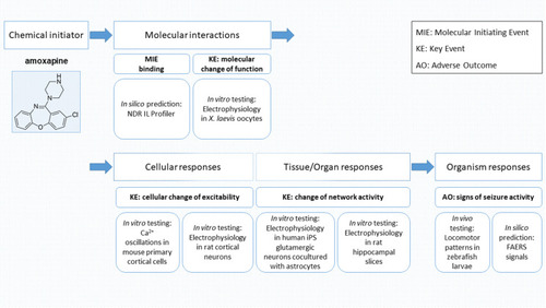
Workflow mapped to adverse outcome pathway (AOP) scales: Assays used for in silico, in vitro, and in vivo experiments to identify molecular initiating events (MIEs) and key events (KEs) related to structural alerts for identifying seizure risk adverse outcomes (AOs)

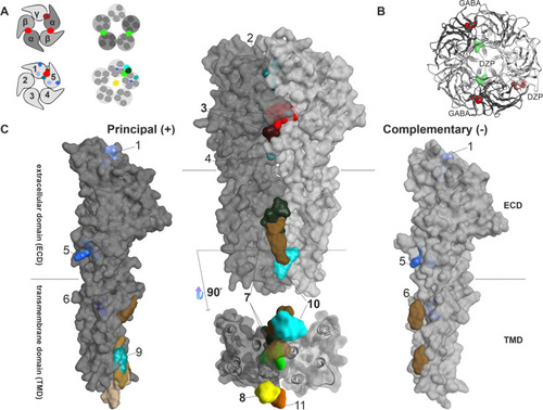
Summary of all identified and putative binding sites, for which structural evidence exists. A Top: representation of αβγ GABAAR pentamers. Below: representation of a generic pentamer. Representative intrasubunit binding sites and interfaces found on the ECD (left), TMD (right), as well as the channel pore (right) are depicted. Colors are matching the ones depicted in panels B, C. B Top view of an atomic model of an α1β3γ2 GABAAR (6HUP) in ribbon representation, with GABA and diazepam binding to the ECD, as well as the diazepam TMD binding site shown in surface filling representation. C All intrasubunit binding sites that can be found on the principal ( +) side in dark grey (left). Representation of all binding sites that occupy an interface between two subunits (middle). Representation of all intrasubunit binding sites that can be found on the complementary (-) side in light grey (right). The extracellular and transmembrane domains are marked as ECD and TMD on the individual subunit renderings, and dashed lines indicate the approximate localization of the lipid collar in the space filling renderings. Below the upright dimer, a view of the transmembrane domain binding sites as seen from the intracellular space is depicted. The surface maps of the principal and complementary sides that are depicted in this summary were obtained from 6HUP. All ligands are shown in space filling representation (representative ligands for each site: 1—“fragment” in sky blue, 2—AM-3607 in turquoise, 3—GABA/diazepam in red and ketamine in dark red, 4—Ba2 + -atom in cadet grey, 5—chlorpromazine in ocean blue, 6—propofol in navy blue, 7—diazepam in light green and avermectin in dark green, 8—picrotoxin in yellow, 9—pregnenolone sulfate in light cyan, 10—alphaxolone in dark cyan, 11—memantine in orange. Brown sites represent lipid-associated sites (e.g., cholesterol in light and dark brown, PIP2 in sand). Sites 1, 5, 6, and 9 are intrasubunit-located, whereas sites 2, 3, 4, 7, and 10 are interface-located. Site 8 and site 11 are located within the channel pore. This summary of binding sites resulted from a superposition of atomic resolution structures of GABAAR and homologous proteins. The PDB files used, citations, as well the full description of the ligands occupying the binding sites can be found in Supplementary Table S1. Note that structural data for the intracellular domain is lacking

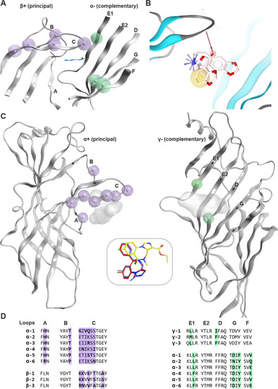
Known ECD interface binding sites for GABA and benzodiazepines: A GABA sites: β3 + / α1 − of 6HUJ showing in lilac residue positions that are different in the beta subunits (β1, β2, β3), as well as those that differ in the alpha subunits in light green. The GABA molecule is rendered in cyan sticks. More details are provided in Supplementary Figure S3. B Chemical feature interactions of the bicuculline bound 6HUK structure rendered with LigandScout 4.4 Expert. Yellow spheres, blue stars, and red vectors represent hydrophobic, positive ionizable, and hydrogen bond acceptor interactions, respectively. C Side view of the benzodiazepine binding site (α + /γ − interface) from a PDBeFold superposition of selected atomic resolution structures (PDB IDs: 6HUP—diazepam, 6HUO—alprazolam, 6D6T/6D6U—flumazenil). The subunits are rendered individually for more clarity, and the variable positions are highlighted as in panel A with lilac for the principal and light green for the complementary face, respectively. The insert box in the middle depicts the binding modes of diazepam (red), alprazolam (blue), and flumazenil (yellow). The corresponding ligands are displayed on the protein as shadows for orientation. The direction of the beta strands on the complementary face is indicated by arrows. D Partial alignment for the binding site forming segments matching panels A and B. More details on variable positions, including those that are found on segment F, are provided in Supplementary Figures S4 and S5)

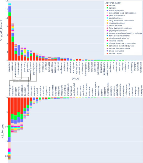
Pharmacovigilance data. The upper box plot displays the fraction (in %) of each AE for a specific drug from the total number of reports for this drug. The drugs are sorted by the total burden of the seizure/convulsion groups of AE, and the AEs in the legend are also sorted by the size of their contribution to the total AE count for these drugs. The lower box plot displays the fraction (in %) of the cumulative seizure AEs for each drug among the total reports in the seizures MedDRA category (46,285 total reports). Compounds which occur in the top 10 of both are connected. The total reports and the reports per AE per drug for the selected drugs and AEs are shown in Supplementary Table S4

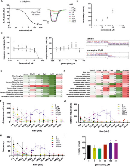
Amoxapine results from different assays. A Dose–response curve of amoxapine derived from TEVC recordings in X. laevis oocytes expressing α1β3γ2 concatenated GABAAR. Representative traces depicted on the right. Data depict mean ± SEM. B Mean (± SEM) concentration–response data summary of amoxapine (3, 10, 30, and 100 μM) effects on CA1 population spike area in rat hippocampal brain slices. Statistical testing was run on raw concentration data (dose: F(4,36) = 22.85, p < .001; Dunnett’s post hoc: amoxapine 30 μM vs veh, p < .05; amoxapine 100 μM vs veh, p < .001). C Calcium oscillation evaluation showing the average of the amoxapine concentration effect on frequency then on amplitude (values depict % of vehicle ratio) and a sample traces recordings for vehicle and amoxapine for the highest amoxapine concentration, namely, 30 µM. D Microelectrode array (MEA) recordings from rat cortical neurons. E Microelectrode array (MEA) recordings with human iPS glutamatergic neurons in co-culture with human astrocytes. The values in the heat maps (D, E) are presented as maximum percentage of change vs baseline after a treatment duration of 1 h (D) or 10 min (E). Each value represents the mean of 3 (D) or 5 (E) wells. A red color shows an increase of the respective endpoint, while a green color shows a decrease of the respective endpoint value compared to baseline (0%). The intensity of the colors indicates the magnitude of the effects as shown below. FI Effect of amoxapine treatment on zebrafish larvae locomotor measurements during 1-h exposure period using video-tracking system (n = 8 larvae/group): F Total distance moved in mm (TDM), median ± median absolute deviation (mad); *p ≤ 0.05, **p ≤ 0.01, and ***p ≤ 0.001 versus control group (2-way ANOVA type with repeated measure on factor time); G total distance moved at high velocity(> 20 mm/s) (TDMH) in mm median ± mad; *p ≤ 0.05, **p ≤ 0.01, and ***p ≤ 0.001 versus control group (2-way ANOVA type with repeated measure on factor time); H frequency at high velocity (> 20 mm/s) (FH), median ± mad; *p ≤ 0.05, **p ≤ 0.01, and ***p ≤ 0.001 versus control group (2-way ANOVA type with repeated measure on factor time); I maximal velocity (MV) in mm/s, median ± mad; **p ≤ 0.01 versus control group (Dunnett’s test after transformation on rank)

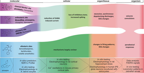
Candidate AOPs for several of the compounds we investigated in this study. The left set of events in blue/ purple hues represent the binding of ligands to their respective binding sites. Picrotoxin, bicuculline, DMCM, clobazam, and amoxapine are rendered in 2D as examples for the different binding sites and for the experimental pipeline. Different allosteric sites can mediate functional agonism and antagonism as well as NAM and PAM effects, which induce typically a change in GABA-elicited current. The proposal reflects a coarse grain model which requires further details to generate complete AOPs. Here, green hues indicate the late molecular and the cellular scales at which the ligand binding leads to changes in inhibitory transmission and then to changes in neuronal firing patterns. The red hues represent the organ and organism scales, at which changed neuronal firing patterns impact on network activity and thus on EEG and ultimately lead to organism responses such as seizures, convulsions, or paradoxical responses such as agitation that are often observed for GABAAR targeting “tranquilizers.” The assays that were used in this study at the molecular, cellular, tissue, and organism scales are integrated at the bottom of the graph. Ligand examples for each pathway are boldfaced and underlined for agents with known seizurogenic properties, boldfaced for strong candidates (meeting at least two criteria), and in standard font for the remaining examples

Acknowledgments
This image is the copyrighted work of the attributed author or publisher, and ZFIN has permission only to display this image to its users. Additional permissions should be obtained from the applicable author or publisher of the image. Full text @ Cell Biol. Toxicol.