|
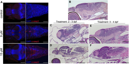
Bilirubin-induced neurological damage (BIND) is detected in the larval brain:(A) Apoptotic cells were detected in the brains of control and hyperbilirubinemia larvae at 4 dpf. DAPI (blue) highlights the brain structure, and cleaved caspase 3 (C-Casp3) (red) labels apoptotic cells. Dorsal views of fore brain (left) and hind brain (right) are shown for control, 9 μM and 15 µM bilirubin treatment groups. (B–F) H&E-stained paraffin sections of 6 dpf larval brains. (A) Intact and healthy brain tissue in control larva; deformed brain tissue in larvae that received (B) 9 µM or (C) 15 µM bilirubin between 2–3 dpf. (D) Larva that received 9 µM bilirubin between 3 - 4 dpf had normal brain tissue. (E) Larva that was exposed to 15 µM bilirubin between 3–4 dpf had slight deformation in brain tissue. Scale bars: 100 µm.
|