
Fig. 6
- ID
- ZDB-FIG-231130-13
- Publication
- Packard et al., 2023 - Zebrafish crocc2 mutants exhibit divergent craniofacial shape, misregulated variability, and aberrant cartilage morphogenesis
- Other Figures
- All Figure Page
- Back to All Figure Page
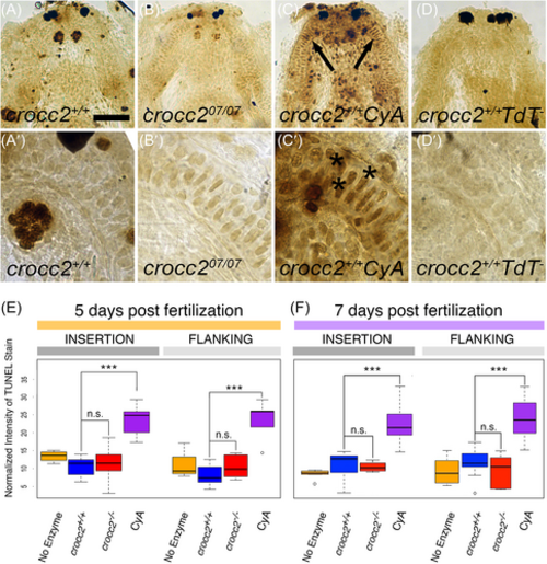
TUNEL staining shows no increase in apoptotic cell death in crocc2 mutant larval cartilages. (A-D) Low and (A′-D′) high magnification ventral views of 7 dpf larvae mandibles showing dark staining in apoptotic cells as detected using the TUNEL method. In wild-type animals (A and A′), a few darkly labeled cells are seen throughout the mandible as compared to the negative control samples where no reaction enzyme was added (TdT−; D and D′). In the positive control samples (CyA; C and C′), wild-type (crocc2+/+) larvae were incubated in cyclopamine (CyA) and show numerous darkly stained cells throughout the mandible. MC cells in crocc207/07 mutant animals (B and B′) exhibit patterns of TUNEL staining similar to what is seen in wild-type siblings. Scale Bar = 50 μm for (A-D) and 10 μm for (A′-D′). (E, F) Box plots displaying the intensity of TUNEL staining, relative to background staining, in individual MC cells adjacent to the muscle insertion and flanking sites. In both (E) 5 dpf larvae and (F) 7 dpf larvae, there are no significant differences (n.s.) in TUNEL staining between wild-type (blue), crocc2 mutant (red), and wild-type negative control (no enzyme, yellow) cartilages and there is a significant increase in TUNEL stain in the positive control samples (CyA = cyclopamine, purple; *** = P < .0001). |