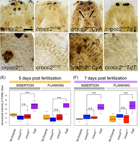
Fig. 6 TUNEL staining shows no increase in apoptotic cell death in crocc2 mutant larval cartilages. (A-D) Low and (A′-D′) high magnification ventral views of 7 dpf larvae mandibles showing dark staining in apoptotic cells as detected using the TUNEL method. In wild-type animals (A and A′), a few darkly labeled cells are seen throughout the mandible as compared to the negative control samples where no reaction enzyme was added (TdT−; D and D′). In the positive control samples (CyA; C and C′), wild-type (crocc2+/+) larvae were incubated in cyclopamine (CyA) and show numerous darkly stained cells throughout the mandible. MC cells in crocc207/07 mutant animals (B and B′) exhibit patterns of TUNEL staining similar to what is seen in wild-type siblings. Scale Bar = 50 μm for (A-D) and 10 μm for (A′-D′). (E, F) Box plots displaying the intensity of TUNEL staining, relative to background staining, in individual MC cells adjacent to the muscle insertion and flanking sites. In both (E) 5 dpf larvae and (F) 7 dpf larvae, there are no significant differences (n.s.) in TUNEL staining between wild-type (blue), crocc2 mutant (red), and wild-type negative control (no enzyme, yellow) cartilages and there is a significant increase in TUNEL stain in the positive control samples (CyA = cyclopamine, purple; *** = P < .0001).
Image
Figure Caption
Acknowledgments
This image is the copyrighted work of the attributed author or publisher, and
ZFIN has permission only to display this image to its users.
Additional permissions should be obtained from the applicable author or publisher of the image.
Full text @ Dev. Dyn.

