
Fig. 4
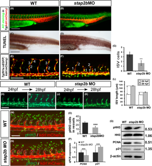
Knockdown of stap2b impairs the growth of ISV. (A–D) AO staining and TUNEL assays were performed to detect apoptotic cells in wild-type control (A and C) and stap2bATG morphants (B and D). Some apoptotic cells were observed in the skin of the morphants, but these cells did not appear at the sites of vasculature. (E and F) Injection of stap2bATG morpholino into Tg (kdrl:mCherry; fli1a:negfp y7) shows the cell number per ISV less than that of the control (M) at 30 hpf. (G) Quantification of average cells per ISV in control and stap2bATG morphants 30 hpf (n = 12 in controls and n = 9 in stap2bATG morphants). (H–K) A migration assay was performed to measure the difference in ISV length from 24 hpf to 28 hpf in control and stap2b morphants (n = 12 in control and morphants), and the quantitative results are shown in (L). (M and N) The proliferation marker pHH3 (green spots) was detected by immunofluorescence in the vessels (red area within white dash line). (O) The number of mitotic cells (pHH3 cells) counted in the controls and stap2b morphants was 25 ± 2.4 and 14.4 ± 2.9, respectively (n = 10 in control and n = 9 in morphants). (P) Quantification of the relative expression level as determined by real-time PCR showed reduced expression of another proliferation marker, PCNA (0.70 ± 0.02), and increased expression of the cell cycle inhibitor p21 (1.59 ± 0.07). (Q) The results from Western blot analysis showed reduced protein expression of pHH3 and PCNA and increased expression of p21. β-actin and histone H3 were the loading controls. The densitometry results are normalized and shown aside. ***Refers to p < .001 and **refers to p < .01 by an unpaired Student's t-test. Scale bars are 200 μm for A–D, and scale bar are 100 μm for E, F, H–K, M and N. |