
Fig. 3
- ID
- ZDB-FIG-231108-68
- Publication
- Tian et al., 2023 - Long-term in toto Imaging of Cellular Behavior During Nerve Injury and Regeneration
- Other Figures
- All Figure Page
- Back to All Figure Page
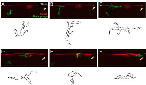
Example of in toto videomicroscopy of axons and macrophages. A–F. Still images from a high-resolution time-lapse recording of a macrophage (green) and a lateralis sensory nerve (red) in the trunk of a larval zebrafish. A single macrophage is seen migrating towards the nerve immediately after it has been severed with a laser. A second macrophage is seen below, which never makes contact with the nerve. Over time, the macrophage moves along the nerve, surveilling damage (which can be seen as a discontinuity of the nerve on the left-hand side of the panels (C and D). The drawings below each panel (dotted lines) describe the two-dimensional shape changes ofthis single macrophage during the course of videomicroscopic imaging. Scale bars is 50 μm. |