
FIGURE
Fig. 1
- ID
- ZDB-FIG-231108-66
- Publication
- Tian et al., 2023 - Long-term in toto Imaging of Cellular Behavior During Nerve Injury and Regeneration
- Other Figures
- All Figure Page
- Back to All Figure Page
Fig. 1
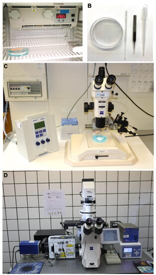
Main tools for the protocol. A. Photograph exemplifying the placement of a plastic Petri dish containing eggs into an incubator. B. Photograph of a standard Petri dish, glass pipette with a hair loop attached to its tip, a forceps and a plastic transfer pipette. C. Example of a stereomicroscope with a typical microinjection apparatus. D. Photograph of an inverted spinning-disc microscope for long-term imaging. |
Expression Data
Expression Detail
Antibody Labeling
Phenotype Data
Phenotype Detail
Acknowledgments
This image is the copyrighted work of the attributed author or publisher, and
ZFIN has permission only to display this image to its users.
Additional permissions should be obtained from the applicable author or publisher of the image.
Full text @ Bio Protoc