
Fig. 4
- ID
- ZDB-FIG-231108-69
- Publication
- Tian et al., 2023 - Long-term in toto Imaging of Cellular Behavior During Nerve Injury and Regeneration
- Other Figures
- All Figure Page
- Back to All Figure Page
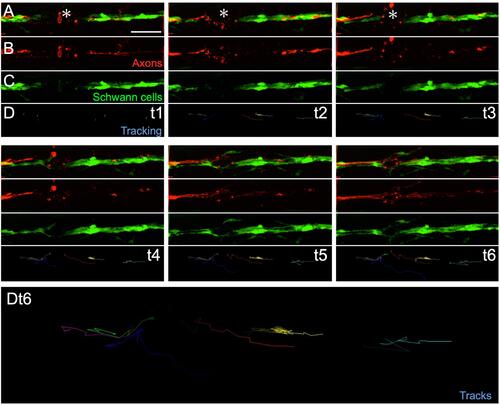
Example of in toto videomicroscopy of axons and Schwann cells. A–C. Fluorescence images from a high-resolution time-lapse recording of the movement of Schwann cells (green) and the regeneration of a lateralis afferent nerve (red) after transection. A shows the merge of the two fluorescence channels, labelling the sensory nerve (Red) and the Schwann cells (green).The respective individual channels are show in B and C. D shows the tracks of the moving Schwann cells. t1, t2, t3, t4, t5, t6 indicate different time points extracted from the time-lapse. Dt6. It a zoom-in of the entire track of Schwann cells of at t6. |