
Fig. 5
- ID
- ZDB-FIG-231106-50
- Publication
- Ding et al., 2023 - Photochemically controlled activation of STING by CAIX-targeting photocaged agonists to suppress tumor cell growth
- Other Figures
- All Figure Page
- Back to All Figure Page
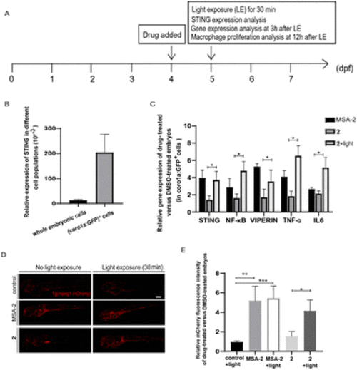
Photo-triggered activation of the STING signalling pathway by 2 in zebrafish. (A) The time scheme for the experiments in (B)–(E). MSA-2 (40 μM) and 2 (10 μM) were added to the embryo medium accordingly. (B) STING is highly expressed in Tg (coro1a:EGFP)+ cells (labelling macrophages and neutrophils). (C) Relative expression of the genes involved in the STING signalling activation in embryos after MSA-2 and 2 treatment with or without light irradiation compared to the DMSO-treated embryos in sorted coro1a:EGFP-labelled cells. Results are expressed as mean ± SEM from three independent experiments, n = 50 embryos per condition (*p < 0.05, t-test). (D) Representative confocal images of Tg (mpeg1:mCherry) embryos (mCherry fluorescence labelling macrophages) after DMSO, MSA-2 and 2 treatment with or without light irradiation. (E) Summary of relative mCherry fluorescent intensity from (D). Results are expressed as mean ± SEM from three independent experiments, n = 15 embryos per condition (***p < 0.001, **p < 0.01, *p < 0.05, t-test). Scale bar in (D): 125 μm; dpf, days post fertilization. |