
Fig. 3
- ID
- ZDB-FIG-231106-48
- Publication
- Ding et al., 2023 - Photochemically controlled activation of STING by CAIX-targeting photocaged agonists to suppress tumor cell growth
- Other Figures
- All Figure Page
- Back to All Figure Page
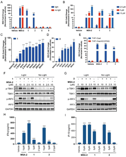
Photo-controllable activation on the STING downstream signalling pathway under blue light. (A) THP1-Dual cells were treated with each compound (MSA-2, 1, 2, 5, and 9) at 10 μM or vehicle (0.1% DMSO), in the presence or absence of blue light (450 nm) for 15 min and then incubated for 24 h. (B) Dose-dependent activation on the ISG reporter of compounds 1 and 2 in THP1-Dual cells under blue light irradiation for 15 min followed by incubation for 24 h. (C) Time-dependent activation on the ISG reporter of compound 1 (5 μM) in THP1-Dual cells under blue light (40 mA, 3.35 mW cm−2) for indicated times, followed by incubation for 24 h. (D) Time-dependent activation on the ISG reporter of compound 2 (5 μM) in THP1-Dual cells under blue light (80 mA, 6.7 mW cm−2) for indicated times, followed by incubation for 24 h. (E) Activation of compounds 1 and 2 (5 μM) on the ISG reporter was dependent on STING under light irradiation for 15 min. The ISG fold change was calculated relative to the vehicle control, and the tests were carried out in triplicate. (F and G) THP1-Dual cells were treated with the indicated concentration of compounds (MSA-2, 1 and 2) with or without 15 min blue light exposure, and then incubated for 4 h. The expression of proteins was determined by western blotting. (H) IFN-β secretion of THP1-Dual cells upon treatment with compounds (MSA-2, 1 and 2) for 24 h after 15 min of blue light exposure. (I) IP-10 secretion of THP1-Dual cells upon treatment with compounds (MSA-2, 1 and 2) for 24 h after 15 min of blue light exposure. Results are expressed as mean ± SEM from two independent experiments. *p < 0.05, **p < 0.01, ***p < 0.001, as compared to the vehicle using one-way ANOVA. |