
Fig. 6
- ID
- ZDB-FIG-231102-21
- Publication
- Zisi et al., 2022 - Small Molecule-mediated Disruption of Ribosome Biogenesis Synergizes With FGFR Inhibitors to Suppress Glioma Cell Growth
- Other Figures
- All Figure Page
- Back to All Figure Page
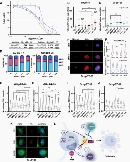
FGFR inhibition potentiates BMH-21 across a pediatric patient-derived cell line panel. (A) BMH-21 dose–response (72 h) in 4 cell lines derived from pediatric GBM tumors with corresponding GI50 values. (B–C) Cas3/7-positive nuclei of GU-pBT-19 and GU-pBT-28 cells. (D) Cell cycle distribution barplot of GU-pBT-7 and GU-pBT-28 following Click-it-EdU-labeling. (E) Representative immunofluorescence images of GU-pBT-28 stained against RPA194&NCL, Scale bar 10 µm (F) RPA194 protein levels quantification by immunofluorescence and RNA synthesis quantified by Click-it-EU-labeling in GU-pBT-28 treated with BMH-21. (G–H) FGF2 and (I–J) SOX2 protein levels quantification by immunofluorescence in GU-pBT-19 and GU-pBT-28 cells. (K) Representative immunofluorescence images of GU-pBT-28 stained against FBL and FGF2, Scale bar 10 µm. The treatment duration was 24 h for (B-F, K) and 48 h for (G–J). (L) Proposed model of synergy: (1) BMH-21 blocks Pol I and downregulates FGFR1 and SOX2., (2) FGF2 is upregulated, released from the nucleolus and secreted, feeding the FGFR1/ERK1/2 feedback loop and rescuing stemness-associated genes to support self-renewal and survival under stress., and (3) Combination of Pol I Inhibition with FGFRi blocks the feedback loop, inhibits the expression of stem cell-associated phenotypes and synergistically induces cell death. Created with Biorender. |