FIGURE

Figure 5—figure supplement 1.

ID
ZDB-FIG-231013-18
Publication
Simon et al., 2023 - Estimating the true stability of the prehydrolytic outward-facing state in an ABC protein
Other Figures
All Figure Page
Back to All Figure Page
Figure 5—figure supplement 1.

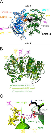
Intra-NBD2 movements associated with ATP binding and tight NBD dimerization.

(A) Location of positions 576, 1370, and 1371 (colored spacefill) relative to composite sites 1 and 2 in the tight hCFTR NBD dimer (PDBID: 6msm). Interacting positions 555 and 1246 (Vergani et al., 2005) are also shown in colored spacefill. (B) NBD2 structures of non-phosphorylated apo IF hCFTR (6uak; light green), non-phosphorylated ATP-bound IF hCFTR (8fzq; medium green), and phosphorylated ATP-bound OF E1371Q-hCFTR (6msm; dark green), aligned through their core subdomains. ATP (shades of yellow), and Mg2+ (shades of purple) of the ATP-bound structures are shown as sticks and spheres, respectively. Note completion of subdomain closure (curved arrow) in the OF structure. (C) Atomic models (sticks and spheres) of ATP (shades of yellow), Mg2+ (shades of purple), and residues D1370 (shades of gray) and S1251 (shades of green) from ATP-bound non-phosphorylated IF WT hCFTR (lighter tones; PDBID: 8fzq) and phosphorylated ATP-bound OF E1371Q hCFTR (darker tones; PDBID: 6msm). The core subdomain of NBD2 from 6msm was aligned with that of 8fzq in Pymol. Residue h1371 (black sticks) is shown only for the OF structure.

Expression Data

Expression Detail
Antibody Labeling
Phenotype Data

Phenotype Detail
Acknowledgments
This image is the copyrighted work of the attributed author or publisher, and ZFIN has permission only to display this image to its users. Additional permissions should be obtained from the applicable author or publisher of the image. Full text @ Elife