|
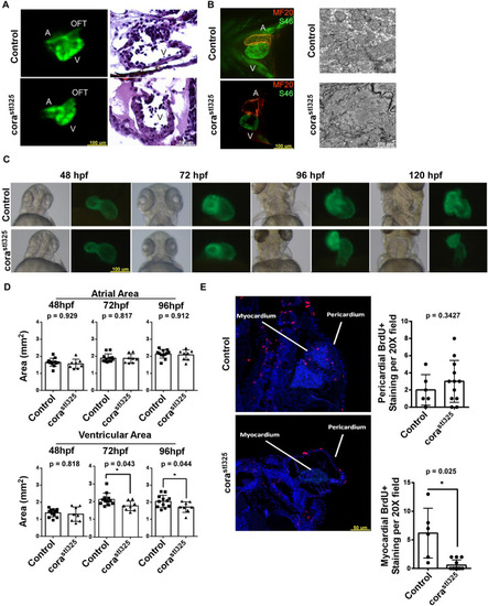
corastl325 hearts display hypoplasia and reduced proliferation. (A) Fluorescent microscopy images of 96 hpf embryos (left, cmlc2::GFP; n=8) and H&E staining (right; n=4). (B) Fluorescent microscopy images of 96 hpf embryos stained with the SF46 (orange) and MF20 (green) showing normal atrioventricular patterning (left). Electron microscopy showing intact sarcomeres in control and corastl325 cardiomyocytes (right; n=4). (C) Brightfield and fluorescence microscopy time-course of cmlc2::GFP expression between 48 and 120 hpf (Control (n=12) and corastl325 (n=8). (D) Quantification of atrial and ventricular cross-sectional areas obtained from C [Control (n=12) and corastl325 (n=8)]. (E) BrdU immunostaining (red) of 96 hpf embryos embedded in paraffin [Control (n=6) and corastl325 (n=12)].
|