
FIGURE
Fig. 1
- ID
- ZDB-FIG-230906-13
- Publication
- Arias-Alpizar et al., 2022 - Phase-separated Liposomes Hijack Endogenous Lipoprotein Transport and Metabolism Pathways to Target Subsets of Endothelial Cells in vivo
- Other Figures
- All Figure Page
- Back to All Figure Page
Fig. 1
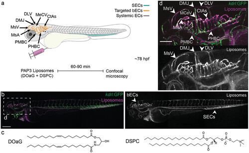
Biodistribution of PAP3 liposomes within zebrafish embryos (78 hpf). a) Schematic zebrafish larvae in lateral (whole-body) view, showing the site of microinjection and key cranial vessels. Fluorescently labeled liposomes are imaged with confocal microscopy after 60–90 min. The vasculature is as follows, liposome targeted bECs in yellow and systemic endothelium in dark gray, scavenger endothelial cells (SECs) in cyan, at ≈78 h post-fertilization (hpf). b) Biodistribution (10x magnification, lateral view) of PAP3 liposomes within a Tg(kdrl:GPF) zebrafish embryo at 1.5 h post-injection (hpi). c) Chemical structure of lipids used in the equimolar mixture for the formulation of PAP3 liposomes, DOaG, and DSPC lipids. d) Zoom of the cranial region in lateral view. bECs, brain endothelial cells; CtAs, central arteries; DLV, dorsal longitudinal vein; DMJ, dorsal midline junction; MCeV, middle cerebral vein; MsA, mesencephalic artery; MsV, mesencephalic vein; PMBC, primordial midbrain channel; PHBC, primordial hindbrain channel; SECs, scavenging endothelial cells. Liposomes formulated by extrusion (5 mM, 0.2% mol DOPE-LR). Scale bars: 200 µm (lateral view) and 100 µm (zoom).
|
Expression Data
Expression Detail
Antibody Labeling
Phenotype Data
Phenotype Detail
Acknowledgments
This image is the copyrighted work of the attributed author or publisher, and
ZFIN has permission only to display this image to its users.
Additional permissions should be obtained from the applicable author or publisher of the image.
Full text @ Adv. Healthc. Mater.