
FIGURE
Fig. 5
- ID
- ZDB-FIG-230906-17
- Publication
- Arias-Alpizar et al., 2022 - Phase-separated Liposomes Hijack Endogenous Lipoprotein Transport and Metabolism Pathways to Target Subsets of Endothelial Cells in vivo
- Other Figures
- All Figure Page
- Back to All Figure Page
Fig. 5
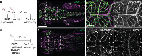
Internalization of PAP3 in zebrafish bECs. a) Timeline of injection and imaging. b,c) Biodistribution (10x and 40x magnification) of PAP3 liposomes (5 mM, 0.2 mol% DOPE-LR) after post-administration of heparin (1 nl, 50 mg ml−1) in a Tg(kdrl:GFP) zebrafish embryo at 1.5 hpi. d) Timeline of injection and imaging. e,f) Biodistribution of PAP3 liposomes (10 mM), containing 0.5 mol% of pH-sensitive DOPE-pHrodo (green/grey) to indicate endocytosis and 0.5 mol% of non-sensitive pH dye DOPE-NBD (magenta/grey) to label liposomes, in an AB/TL zebrafish embryo at 1.5 hpi. All zebrafish larvae at ≈78 hpi. Liposomes formulated by extrusion. Scale bars: 200 µm (whole embryo) and 50 µm (tissue level).
|
Expression Data
Expression Detail
Antibody Labeling
Phenotype Data
Phenotype Detail
Acknowledgments
This image is the copyrighted work of the attributed author or publisher, and
ZFIN has permission only to display this image to its users.
Additional permissions should be obtained from the applicable author or publisher of the image.
Full text @ Adv. Healthc. Mater.