
Fig. 2
- ID
- ZDB-FIG-230831-14
- Publication
- Jackson et al., 2023 - Clinical, genetic, epidemiologic, evolutionary, and functional delineation of TSPEAR-related autosomal recessive ectodermal dysplasia 14
- Other Figures
- All Figure Page
- Back to All Figure Page
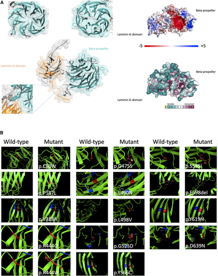
In silico protein modeling of pathogenic/likely pathogenic non-truncating TSPEAR variants (A) Left shows the AlphaFold-predicted structure of TSPEAR with all missense/in-frame indels plotted; note the propensity for these to affect the β-propeller. Right shows the surface of the predicted TSPEAR structure showing overall charge. Note the pocket of negative charge (red) within the inner surface of the β-propeller. Conservation scores are shown below mapped to the predicted structure surface showing conservation of the residues (purple) within the inner surface of the β-propeller. (B) Molecular models for all missense/in-frame indels in this study produced by Modeller 9.24. Steric clashes are shown by red discs. Yellow dotted lines indicated interacting residues. |