Fig. 6
- ID
- ZDB-FIG-230714-27
- Publication
- Chen et al., 2022 - Increasing the field-of-view in oblique plane microscopy via optical tiling
- Other Figures
- All Figure Page
- Back to All Figure Page
|
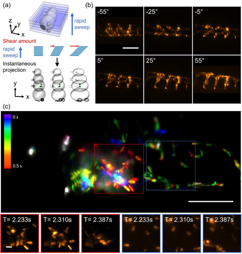
Extending the field of view in real time projection imaging. (a) Schematic representation of the projection method: the light-sheet is rapidly swept through the sample, and the instantaneous images are translated at the same rate on the camera sensor. Depending on the amount of lateral translation, projections under different viewing angle are obtained. (b) Projections of vasculature in the zebrafish tail under different viewing angles. (c) Tiled projection imaging of fluorescently labeled blood cells in a zebrafish heart. Insets show a montage of blood cells at different timepoints. Scale bars: (b-c) 100 µm and insets 10 µm. |

