
FIGURE
Fig. 1
- ID
- ZDB-FIG-230714-22
- Publication
- Chen et al., 2022 - Increasing the field-of-view in oblique plane microscopy via optical tiling
- Other Figures
- All Figure Page
- Back to All Figure Page
Fig. 1
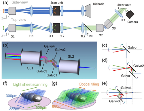
(a) Schematic drawing of the OPM system with optical tiling. O1-3, objectives; TL1-3, Tube Lenses; SL1-2, Scan Lenses, IM, Illumination Module. (b) Rendering of the optical layout of the dual-axis scan unit and scan lenses. (c)-(e) Working principle of a single-galvo, dual-galvo and quad-galvo scan unit. (f) Image volume covered by conventional OPM (blue volume). (g) Image volume covered by optical tiling OPM (blue and green volumes). The pink circle depicts the field of view of the primary objective. |
Expression Data
Expression Detail
Antibody Labeling
Phenotype Data
Phenotype Detail
Acknowledgments
This image is the copyrighted work of the attributed author or publisher, and
ZFIN has permission only to display this image to its users.
Additional permissions should be obtained from the applicable author or publisher of the image.
Full text @ Biomed. Opt. Express