FIGURE
Fig. 2
- ID
- ZDB-FIG-230714-23
- Publication
- Chen et al., 2022 - Increasing the field-of-view in oblique plane microscopy via optical tiling
- Other Figures
- All Figure Page
- Back to All Figure Page
Fig. 2
|
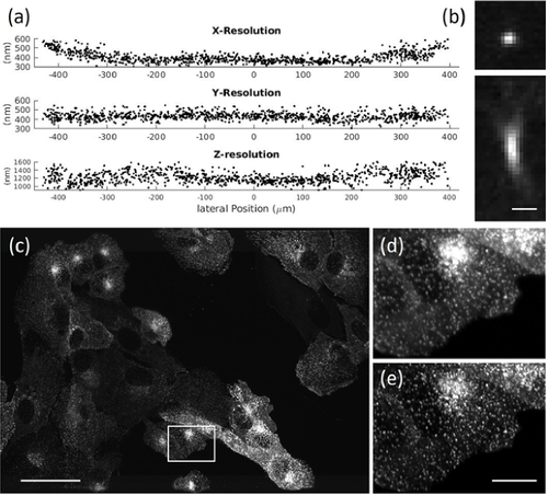
(a) Full Width Half Maximum measurements of fluorescent nanospheres over the whole FOV. (b) Representative point-spread function of a fluorescent nanosphere. (c) Maximum intensity projection of parental human retinal pigmented epithelium (ARPE-19) cells EGFP-labeled for AP2. (d) Raw data for box in (c). (e) Deconvolved data for box in (c). Scale bars: (b)1 µm; (c) 50 µm; (e) 10 µm. |
Expression Data
Expression Detail
Antibody Labeling
Phenotype Data
Phenotype Detail
Acknowledgments
This image is the copyrighted work of the attributed author or publisher, and
ZFIN has permission only to display this image to its users.
Additional permissions should be obtained from the applicable author or publisher of the image.
Full text @ Biomed. Opt. Express

