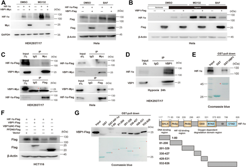
VBP1 interacts with HIF-1α and induces HIF-1α degradation.A, The downregulation of HIF-1α protein caused by ectopic expression of VBP1 was blocked by MG132 and BAF. Cells were transfected with the indicated plasmids and harvested 24 h after transfection; MG132 (left panel) or BAF (right panel) were added to the culture medium at 16 h after transfection. and the proteins were detected by Western blotting analysis. (−), the cells transfected with the empty vector control; (+), the cells transfected with the indicated vector. B, The downregulation of endogenous HIF-1α protein caused by ectopic expression of VBP1 in the hypoxic cells could be rescued by treatment with MG132 and BAF. HeLa cells transfected with VBP1-Flag or a control plasmid were incubated under normoxia or hypoxia for 24 h, and treated with vehicle or 10 μM MG132 or 50 nM BAF for 8 h. Cell lysates were immunoblotted for HIF-1α. C, VBP1 interacts with HIF-1α. The interaction between VBP1-Myc and Flag-HIF-1α was analyzed in HEK293T/17 cells (left panel) and HeLa cells (right panel) by reciprocal Co-IP as indicated. D, Endogenous VBP1 interacts with HIF-1α in HEK293T/17 cells. Anti-VBP1 antibody was used for IP. E, GST pull-down assay results show the interaction between HIF-1α and GST-tagged VBP1. F, Overexpression of VBP1ΔNC resulted in a decrease of HIF-1α. HCT116 cells were transfected with the indicated plasmids and harvested after transfection of 24 h and the proteins were detected by Western blotting analysis. G, Mapping the interaction domain of HIF-1α with VBP1. GST pull-down assays were performed with GST or GST fusion proteins containing the indicated amino acid residues of HIF-1α (top) and the whole-cell lysates of HEK293T/17 cells expressing VBP1-Flag. bHLH, basic helix-loop-helix; HIF-1, Hypoxia-inducible factor-1; PAS, Per-ARNT-Sim; pVHL, von Hippel-Lindau protein; TAD, transactivation domain; VBP1, pVHL binding protein 1.
|
