FIGURE SUMMARY
Title

VBP1 negatively regulates CHIP and selectively inhibits the activity of hypoxia-inducible factor (HIF)-1α but not HIF-2α

Authors
Yue, Y., Tang, Y., Huang, H., Zheng, D., Liu, C., Zhang, H., Liu, Y., Li, Y., Sun, X., Lu, L.
Source
Full text @ J. Biol. Chem.

Generation of the transgenic HRE-sv40mp:GFP reporter zebrafish.A, Schematic representation of the Tol-2 vector used to generate the Tg (hre-sv40mp: GFP) zebrafish. The construct includes a 68-bp fragment from the hypoxia-response element (HRE) of the human ENO1 gene and the SV40 minimal promoter (SV40mp). B, Representative images show the Tg (hre-sv40mp:GFP) embryos at 48 and 72 hpf under normoxic or hypoxic conditions. Tg (hre-sv40mp:GFP) embryos were exposed to hypoxia (10% O2) for 24 or 48 h from 24 hpf. Transgenic embryos show strong GFP expression during hypoxia. However, GFP expression is observed only in the visceral organs in the transgenic embryos exposed to normoxia. C, Tg(hre-sv40mp:GFP) embryos injected with the dominant-active (DA) HIF-1α mRNA at the 1-cell stage show strong GFP expression at 48 hpf. D, Transgenic zebrafish in the vhl−/− background show strong GFP fluorescence compared to the sibling embryos at 72 hpf. Scale bar, 500 μm.

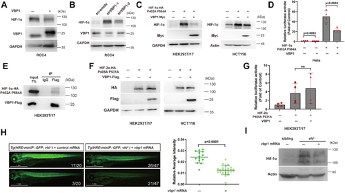
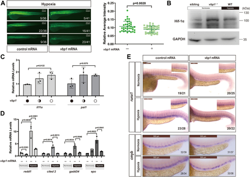
Vbp1 negatively regulates the HIF-1α signaling pathway in zebrafish.A, Representative images show GFP fluorescence in the Tg (hre-sv40mp:GFP) embryos that were injected with mCherry or vbp1 mRNA at the 1-cell stage and exposed to hypoxia for 48 h from 24 hpf. The histograms show average GFP fluorescence intensity in the somite of the Tg(hre-sv40mp:GFP) larvae (n ≥ 36) at 72 hpf. B, Western blot shows Hif-1α protein levels in the sibling, vbp1−/−, wt (hypoxia treatment) larvae at four dpf. As shown, Hif-1α is upregulated in the vbp1 mutants. C, The relative mRNA levels of hypoxia-responsive genes, namely, il11a, and pai1 in the WT, vbp1+/−, and vbp1−/− larvae. The data show upregulation of these hypoxia-responsive genes in the vbp1 mutant zebrafish (n = 3, biologically independent extracts). D, Effect of forced expression of vbp1 on redd1, cited2, gadd34, and epo mRNA expression. Embryos injected with GFP or vbp1 capped mRNA was raised to 24 hpf and then exposed to hypoxia (10% O2) for 24 h. The mRNA levels of the indicated genes were determined by qPCR. (n = 3, biologically independent extracts). E, Representative images show both runx1 and cmyb expression in the zebrafish embryos injected with control or vbp1 mRNAs under normoxic and hypoxic conditions. As shown, runx1/cmyb expression is reduced in the vbp1-injected embryos under hypoxia conditions. The brightfield images of WISH for runx1/cmyb expression are shown in the WT embryos at 36 hpf under normoxia and hypoxia exposure for 8 h starting at 28 hpf (lateral views). Scale bar, 250 μm and 500 μm representatively. All data are represented as means ± SD. n/n, number of embryos showing representative phenotype/total number of embryos examined. HIF-1, Hypoxia-inducible factor-1; pVHL, von Hippel-Lindau protein; VBP1, pVHL binding protein 1.

VBP1 negatively regulates HIF-1α but not HIF-2α.A, Western blot shows protein levels of HIF-1α but not HIF-2α were elevated in VBP1 knockdown HCT116 cells. The cells were stably transfected with the lentiviral pLKO vector containing specific shRNAs targeting VBP1 or control scrambled shRNA and exposed to either 21% or 1% O2 for 6 h. B, HIF-1α protein levels were downregulated under hypoxic conditions. Western blotting analysis of control and VBP1-overexpressing HCT116 cells cultured at the indicated oxygen levels for 24 h. CE, Overexpression of VBP1 decreased the HIF-1α but not HIF-2α protein levels. HCT116, U2OS, and HEK293T/17 cells were transfected with the indicated plasmids, and the proteins were detected by Western blotting analysis. (−), the cells transfected with the empty vector control; (+), the cells transfected with the indicated vector. F, Overexpression of VHL decreased HIF-2α protein levels but VBP1 failed to decrease it. 786-O cells were transfected with the indicated plasmids, and the proteins were detected by Western blotting analysis. G, ENO1 promotor-luciferase reporter activity in the HEK293T/17 and the HeLa cells show that VBP1 decreases HIF-1α but not HIF-2α transcriptional activity. The firefly luciferase activities were normalized to the Renilla luciferase activity. n = 3 biological replicate samples. H, VEGF promotor-luciferase reporter activity in the HEK293T/17 and the 786-O cells show that VBP1 failed to decrease HIF-2α or HIF-2α (P405A/P531A) transcriptional activity. The firefly luciferase activities were normalized to the Renilla luciferase activity. n = 3 biological replicate samples. I, RT-qPCR analysis of HIF target gene induction in response to hypoxia in HEK293T/17 cells transfected with empty vector or Flag-tagged VBP1 vector. The data are represented as means ± SD. n = 3 biologically independent extracts. ns, not significant. HIF-1, Hypoxia-inducible factor-1; pVHL, von Hippel-Lindau protein; VBP1, pVHL binding protein 1.

VBP1 interacts with HIF-1α and induces HIF-1α degradation.A, The downregulation of HIF-1α protein caused by ectopic expression of VBP1 was blocked by MG132 and BAF. Cells were transfected with the indicated plasmids and harvested 24 h after transfection; MG132 (left panel) or BAF (right panel) were added to the culture medium at 16 h after transfection. and the proteins were detected by Western blotting analysis. (−), the cells transfected with the empty vector control; (+), the cells transfected with the indicated vector. B, The downregulation of endogenous HIF-1α protein caused by ectopic expression of VBP1 in the hypoxic cells could be rescued by treatment with MG132 and BAF. HeLa cells transfected with VBP1-Flag or a control plasmid were incubated under normoxia or hypoxia for 24 h, and treated with vehicle or 10 μM MG132 or 50 nM BAF for 8 h. Cell lysates were immunoblotted for HIF-1α. C, VBP1 interacts with HIF-1α. The interaction between VBP1-Myc and Flag-HIF-1α was analyzed in HEK293T/17 cells (left panel) and HeLa cells (right panel) by reciprocal Co-IP as indicated. D, Endogenous VBP1 interacts with HIF-1α in HEK293T/17 cells. Anti-VBP1 antibody was used for IP. E, GST pull-down assay results show the interaction between HIF-1α and GST-tagged VBP1. F, Overexpression of VBP1ΔNC resulted in a decrease of HIF-1α. HCT116 cells were transfected with the indicated plasmids and harvested after transfection of 24 h and the proteins were detected by Western blotting analysis. G, Mapping the interaction domain of HIF-1α with VBP1. GST pull-down assays were performed with GST or GST fusion proteins containing the indicated amino acid residues of HIF-1α (top) and the whole-cell lysates of HEK293T/17 cells expressing VBP1-Flag. bHLH, basic helix-loop-helix; HIF-1, Hypoxia-inducible factor-1; PAS, Per-ARNT-Sim; pVHL, von Hippel-Lindau protein; TAD, transactivation domain; VBP1, pVHL binding protein 1.

VBP1 negatively modulates HIF-1α protein levels in a pVHL-independent manner.A, Overexpression of VBP1 decreased the HIF-1α protein levels in pVHL-deficient RCC4 cells. RCC4 cells were stably transfected with the control or pCS2-VBP1 plasmid, and the proteins were detected by Western blotting analysis. B, Protein levels of HIF-1α were elevated in VBP1 knockdown RCC4 cell lines. Immunoblot analysis of cells stably transfected with lentiviral pLKO plasmid with VBP1-specific shRNA or scrambled control shRNA. C, VBP1 decreases HIF-1α (P402A/P564A) protein levels. HEK293T/17 and HCT116 cells were transfected with the indicated plasmids, and the proteins were detected by Western blotting analysis. D, HIF luciferase reporter assay results in HeLa cells show that VBP1 decreased HIF-1α (P402A/P564A) transcriptional activity. Data are represented as means ± SD (n > 3 biological replicate samples). E, VBP1 interacts with HIF-1α (P402A/P564A). Co-IP was performed with lysates from HEK293T/17 cells co-transfected with plasmids overexpressing VBP1-Flag and HA-tagged HIF-1α (P402A/P564A). F, VBP1 has no effect on the protein levels of HIF-2α (P405A/P531A). Immunoblot analysis of cells transfected with the indicated plasmids. G, HIF luciferase reporter assay results in HEK293T/17 cells show that VBP1 does not affect the HIF-2α (P405A/P531A) transcriptional activity. ns, not significant. n > 3 biological replicate samples. H, Vbp1 negatively modulates the HIF-1α signaling pathway in Tg (hre-sv40mp:GFP) zebrafish in the vhl−/− background. Tg(hre-sv40mp:GFP) embryos were injected with control mRNA or vbp1 mRNA, and images were taken in 72 hpf. The histograms show the average values (means ± SD) of GFP intensity in the somite of the Tg(hre-sv40mp:GFP) vhl−/− larvae at 72 hpf. n/n, number of embryos showing representative GFP fluorenes/total number of embryos examined. I, Vbp1 negatively modulates the Hif-1α protein levels in vhl−/− zebrafish. Zebrafish embryos injected with control mRNA or vbp1 mRNA were collected at 72 hpf and the proteins were detected by Western blotting analysis. HIF-1, Hypoxia-inducible factor-1; pVHL, von Hippel-Lindau protein; VBP1, pVHL binding protein 1.

CHIP is required for VBP1-mediated HIF-1α stabilization.A, Interaction between VBP1-Flag and HSP70-HA was analyzed in the HEK293T/17 cells by reciprocal Co-IP as indicated. B, Endogenous HSP70 and CHIP interact with VBP1 in the HEK293T/17 cells. Anti-VBP1 antibody was used for Co-IP. C, Interaction between VBP1-Flag and CHIP-HA was analyzed in the HEK293T/17 cells by reciprocal Co-IP as indicated. D, Overexpression of VBP1 increased the CHIP protein levels. HeLa cells were transfected with the indicated plasmids, and the proteins were detected by Western blotting analysis. E, VBP1 increased the endogenous CHIP protein levels. HeLa cells were transfected with VBP1-Myc, and the proteins were detected by Western blotting analysis. F, Western blotting analysis shows that CHIP protein levels were reduced in the VBP1-silenced HEK293T/17 cell lines. The HEK293T/17 cells were stably transfected with lentiviral pLKO plasmids cloned with specific shRNA targeting VBP1 or control scrambled shRNA. G, VBP1-Flag expression decreased CHIP protein half-life. HeLa cells with ectopic VBP1-Flag expression or control plasmids were treated with 50 μg/ml CHX for indicated time periods before being collected for western blotting analysis. The means and SDs from three independent experiments are shown (lower panel). H, Knockdown CHIP diminished the inhibitory effect on ectopic overexpressed HIF-1α by VBP1 in the HCT116 cells. Cells were transfected with the indicated plasmids, and the proteins were detected by Western blotting analysis. The bands of Flag-HIF-1α (∼110 Kd) and VBP1-Flag (∼25 Kd) were easily distinguished by molecular weight. I, Knockdown CHIP diminished the inhibitory effect on endogenous HIF-1α by VBP1 in cells. HCT116 cells transfected with VBP1-Flag or control plasmids were incubated under normoxia or hypoxia for 12 h, and the proteins were detected by Western blotting analysis. J, HEK293T/17 cells with or without silencing of CHIP expression were transfected with the indicated plasmids and treated with MG132 for 8 h. Cell lysates were immunoprecipitated with anti-Flag antibody. The immunoprecipitates and input were probed for indicated antibodies by immunoblotting. K, Mapping the interaction domain of CHIP with VBP1. GST pull-down assays were performed with the GST or GST fusion proteins containing different domains of the CHIP protein. The whole-cell lysates were prepared from HEK293T/17 cells expressing the Flag-tagged VBP1 protein. HIF-1, Hypoxia-inducible factor-1; HSP, heat shock protein; pVHL, von Hippel-Lindau protein; VBP1, pVHL binding protein 1.

VBP1 expression is associated with the overall survival of ccRCC patients.A, Kaplan-Meier survival curves revealed that patients with low levels of VBP1 (bottom 50%, TPM < 5.94633) had significantly lower overall survival in the ccRCC, n = 532. B, Patients with mutated VHL with low levels of VBP1 (TPM < 5.94633) had significantly lower overall survival in the ccRCC, n = 163. RNA-seq expression profiles of VBP1 gene and corresponding clinical information for ccRCC and ccRCC VHL mutant patients were obtained from TCGA database. All data were standard TPM data and the statistical analyses were performed using R software. ccRCC, clear cell renal carcinoma; OS, overall survival; VBP1, pVHL binding protein 1.

Acknowledgments
This image is the copyrighted work of the attributed author or publisher, and ZFIN has permission only to display this image to its users. Additional permissions should be obtained from the applicable author or publisher of the image. Full text @ J. Biol. Chem.