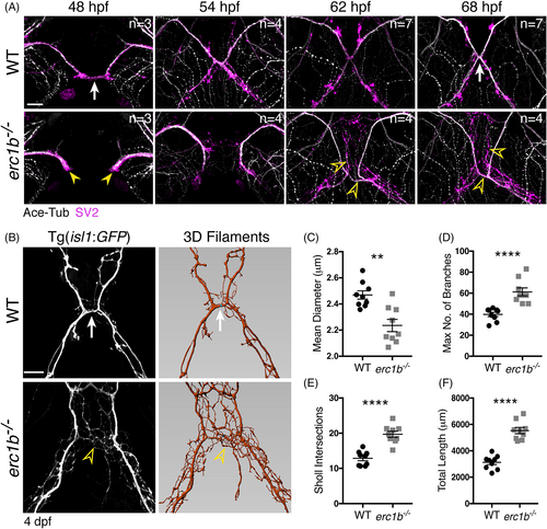
Nerve outgrowth and branching are significantly altered in Erc1b deficient zebrafish. A, Whole-mount IF with antibody labeling acetylated tubulin (white) and SV2 (magenta) in WT and erc1b−/− embryos. White arrow indicates midline contact in WT. Yellow arrowheads indicate delayed trigeminal outgrowth in erc1b−/− embryos. Open yellow arrowheads indicate increased nerve branching in erc1b−/− embryos. Number of examined animals (n) listed in top right of each image. B, Left: images of live transgenic (Tg(isl1:GFP)) WT and erc1b−/− larvae. White arrow indicates nerve branching in WT. Open yellow arrowhead indicates ectopic branching in erc1b−/− larvae. Right: Imaris 3D filament tracing of cranial nerves from images on the left. Quantification of mean diameter of nerve filaments, C, number of filament branches, D, Sholl intersections, E, and total nerve length, F in erc1b−/− and WT larvae. Symbols indicate number of animals (WT n = 9, erc1b−/− n = 9), lines indicate mean with SEM. Mann-Whitney U test (two-tailed) was used for statistical analysis, 95% confidence interval, **P < .01, ****P < .0001. All images are maximum intensity projections, ventral view with anterior on top. Scale bars = 25 μm
|
