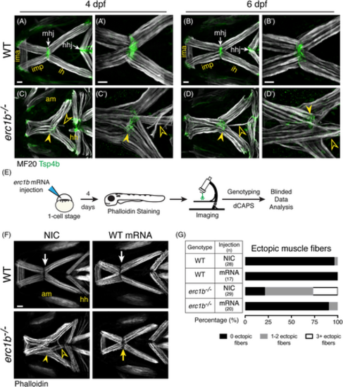
Overexpression of Erc1b rescues ectopic jaw muscle fiber phenotype in Erc1b deficient larvae. Labeling of WT lower jaw muscle fibers with muscle myosin antibody (MF20, white) and myotendinous junction marker (Tsp4b, green, white arrows) at 4 dpf (A [n = 6]) and 6 dpf (B [n = 10]). erc1b−/− larvae at 4 dpf (C [n = 5]) and 6 dpf (D [n = 10]) display partially disorganized lower jaw musculature, including disorganized mhj (closed arrowheads) and 1 to 2 ectopic muscle fibers extending toward the hhj (open arrowheads). A′-D′, Higher magnification images of mhj in individual larvae show severe (3+) ectopic fibers in erc1b−/− larvae, C′-D′. E, Experimental design for zebrafish WT erc1b mRNA injections and subsequent analysis of muscle fiber phenotype. F, Representative images of phalloidin-stained muscles in WT (top) and erc1b−/− (bottom) (4 dpf). NIC erc1b−/− larvae display disorganized mhj (yellow arrowhead) and ectopic muscle fibers (open yellow arrowheads), which are rescued in WT mRNA injected erc1b−/− larvae (yellow arrow). G, Quantification of percent of larvae displaying ectopic muscle fibers. Sample sizes (n) for each genotype and injection denoted in the table. All images are maximum intensity projections; ventral view, anterior to the left. Scale bars = 25 μm. AM, adductor mandibulae; HH, hyohyal; HHJ, hyohyal junction; IH, interhyal; IMA, intermandibularis anterior; IMP, intermandibularis posterior; MHJ, mandibulohyoid junction
|
