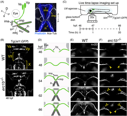
Erc1b deficiency perturbs trigeminal nerve growth. A, Left: schematic of trigeminal motor nerve (Va: anterior trigeminal cluster; Vp: posterior trigeminal cluster) projections into the first (BA1) and second branchial arches (BA2) of zebrafish larvae; IMA: intermandibularis anterior; IMP: intermandibularis posterior; IH: interhyal; MHJ: mandibulohyoid junction. Right: Whole-mount IF of cranial motor nerves (Ace-Tub: acetylated tubulin, white) and lower jaw muscles (phalloidin, blue). Orientation (dorsal (D), ventral (V), anterior (A), posterior (P)) indicated. B, Clusters of cranial motor neurons (Va, Vp: trigeminal; VII: facial; X: vagus) in hindbrain of 48 hours post-fertilization (hpf) WT and erc1b−/− Tg(isl1:GFP) embryos (dorsal view). C, Experimental design for time-lapse imaging of growing trigeminal nerves in transgenic WT and erc1b−/− embryos. Z-stack images were taken every 15 minutes over a 20-hour recording period. D, Schematic of WT trigeminal nerve growth and appearance of lower jaw muscles. E, Stills from time-lapse of trigeminal nerve growth in a WT embryo (ventral view). White arrow indicates midline contact. F, erc1b−/− embryo displays growth delay (yellow arrowheads) and ectopic branching off the anterior side of the trigeminal nerve (open yellow arrowheads). Number of examined animals (n) listed in the first time-lapse images. All images are maximum intensity projections, anterior on top. Scale bars = 25 μm
|
