
Fig. 5
- ID
- ZDB-FIG-230501-172
- Publication
- Yang et al., 2022 - Heterogeneities of zebrafish vasculature development studied by a high throughput light-sheet flow imaging system
- Other Figures
- All Figure Page
- Back to All Figure Page
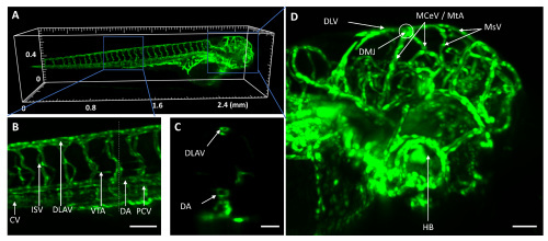
Whole-larva 3D image of zebrafish vasculature captured with LS-FIS at 5 dpf. (A) 3D rendering of the whole larva. See also Visualization 3. (B) Zoomed-in of the box in (A) showing vessels at the truck. CV: caudal vein; ISV: intersegmental vessels; DLAV: dorsal longitudinal anastomotic vessel; VTA: vertebral artery; DA: dorsal aorta; PCV: posterior cardinal vein. (C) Sectioned view along the dashed line of (B). (D) Zoomed-in of the box in (A) showing the vessels at the head. DLV: dorsal longitudinal vein; MCeV: middle cerebral vein; MsV: mesencephalic vein; HB: hyaloid basket. Scale bars represent sent 100 µm for (B, D) and 50 µm for (C), respectively. |