
Fig 7
- ID
- ZDB-FIG-230423-7
- Publication
- Bolten et al., 2023 - Zebrafish (Danio rerio) larvae as a predictive model to study gentamicin-induced structural alterations of the kidney
- Other Figures
- All Figure Page
- Back to All Figure Page
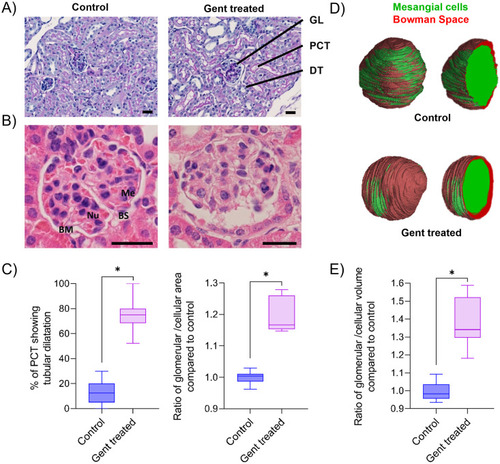
2D and 3D assessment of kidney damage in gentamicin-treated mice.
(A) Representative PAS-stained histology slices (3 μm) from a kidney biopsy of a control and a gentamicin-treated mouse. Gentamicin treatment: daily i.p. injections for ten days of 150 mg gentamicin. Glomerulus = GL, proximal convoluted tubules = PCT, distal tubules = DT. (B) Representative images of an H&E stained 3 μm histological slice of a glomerulus of control and a gentamicin-treated mouse. Mesangial cells = Me, Nuclei = Nu, basal membrane = BM, basal space = BS. (C) Semiquantitative analysis of % of PCT showing tubular dilatation and the ratio of glomerular to cellular area normalized to the mean of the control. 180 PCT of three mice (control or treated) and 30 glomeruli of six mice (control or treated) were analyzed, respectively. (D) SRμCT analysis and visualization of a representative 3D rendered glomerulus of a control and a gentamicin-treated mouse. Mesangial cells are labelled in green, and Bowman’s space in red. (E) SRμCT-based analysis of the ratio of glomerular to cellular volume as compared to the mean of the control. Box plots are shown of five manually segmented glomeruli of three mice each. *p < 0.01. Scale bar: 25 μm. |