
FIGURE
Fig 4
- ID
- ZDB-FIG-230423-4
- Publication
- Bolten et al., 2023 - Zebrafish (Danio rerio) larvae as a predictive model to study gentamicin-induced structural alterations of the kidney
- Other Figures
- All Figure Page
- Back to All Figure Page
Fig 4
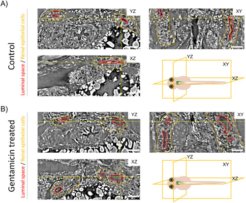
Microtomography-based visualization of the ZFL pronephros.
(A) Orthogonal sections from the 4 dpf ZFL through the microtomography volume of PBS (control) and treated with 1 nL of 42 mM gentamicin (24 hour incubation). Yellow lines show the position of the virtual slices, which intersect at the reticle point located in the luminal space. Luminal space is outlined in red, renal epithelial cells are in yellow. Schematic illustrations indicate the cutting planes. Scale bar: 100 μm. |
Expression Data
Expression Detail
Antibody Labeling
Phenotype Data
Phenotype Detail
Acknowledgments
This image is the copyrighted work of the attributed author or publisher, and
ZFIN has permission only to display this image to its users.
Additional permissions should be obtained from the applicable author or publisher of the image.
Full text @ PLoS One