Figure 1
- ID
- ZDB-FIG-230407-29
- Publication
- Yang et al., 2023 - Release of STK24/25 suppression on MEKK3 signaling in endothelial cells confers Cerebral cavernous malformation
- Other Figures
- All Figure Page
- Back to All Figure Page
|
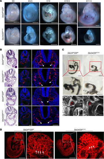
Deletion of Stk24 and Stk25 in endothelium results in vascular defects during embryonic development.
(A) Stereomicroscopic images of developmental time course of littermate Stk24fl/fl;Stk25fl/fl and Stk24/25dECKO mice. Scale bars: 1 mm. (B) H&E staining and Pecam immunostaining of transverse sections of E10 Stk24fl/fl;Stk25fl/fl (n = 3) and Stk24/25dECKO (n = 4) embryos reveal the presence of normally lumenized dorsal aortas (DA) in the Stk24fl/fl;Stk25fl/fl embryos but not in Stk24/25dECKO embryos. White arrowheads indicate dorsal aortas. Scale bars: 100 μm. (C) Images of E9.5 embryo hearts of Stk24fl/fl;Stk25fl/fl (n = 9) and Stk24/25dECKO (n = 6) embryos with injection of Indian ink. Upper and middle panels show embryo overview and magnification of the boxed regions, showing injected ink flows primarily through the second and third BAA to fill the DA in the Stk24fl/fl;Stk25fl/fl embryos. In contrast, ink injected into the heart of Stk24/25dECKO embryos failed to opacify the DA. Accumulation of ink was observed in the heart due to the narrow BAA. Scale bars: 500 μm. Lower panel shows whole-mount immunostaining for the endothelial cell marker, endoglin, showing narrowed BAA and adjacent DA (red arrows) in Stk24/25dECKO embryos. Scale bars: 100μm. (D) Whole-mount immunostaining with endoglin showing the impaired vascular patterning (indicated by the white arrows) in the brain of E9.5 Stk24/25dECKO (n = 6) embryos in comparison with that of Stk24fl/fl;Stk25fl/fl (n = 3) littermate control embryos. Scale bars: 500 μm in overview panels and 100 μm in magnified panels. All the images presented are representatives of 3 or more independent experiments. |

