
Figure 7
- ID
- ZDB-FIG-230406-60
- Publication
- Li et al., 2023 - Copper overload impairs hematopoietic stem and progenitor cell proliferation via prompting HSF1/SP1 aggregation and the subsequently downregulating FOXM1-Cytoskeleton axis
- Other Figures
- All Figure Page
- Back to All Figure Page
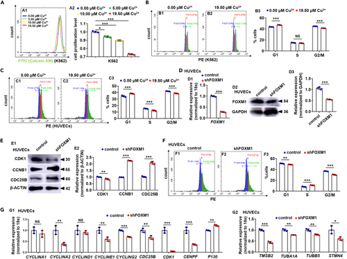
Cu overload impairs the cell cycle in mammalian cells (A) Analysis of cell proliferation in the 0.00 μM Cu, 5.00 μM Cu, 10.00 μM Cu, and 19.50 μM Cu-stressed K562 cells. A1, flow cytometry (FACS) histogram; A2, calculation of cell proliferation in different groups. (B) Cell cycle in 19.50 μM Cu-stressed K562 cells. B1, B2, flow cytometry (FACS) histogram; B3, calculation of cell cycle stage. (C) Cell cycle in 19.50 μM Cu-stressed HUVECs cells. C1, C2, flow cytometry (FACS) histogram; C3, calculation of cell cycle stage. (D) FOXM1 mRNA (D1) and the protein level (D2-D3) were decreased in FOXM1 knockdown (shFOXM1) HUVECs, GAPDH was used as an internal control. (E) Protein levels of cell cycle regulators CDK1, CCNB1, and CDC25B in FOXM1 knockdown (shFOXM1) HUVECs (E1), GAPDH was used as an internal control. (F) shFOXM1 HUVEC cells were blocked at the G1 stage. F1, F2, flow cytometry (FACS) histogram; F3, cell cycle stage calculation. (G) Expression of cell cycle genes (G1) and cytoskeleton genes (G2) in shFOXM1 cells. Data are mean ± SD. t-test, ∗p < 0.05, ∗∗p < 0.01, ∗∗∗p < 0.001. NS, not significant. |