
Figure 3
- ID
- ZDB-FIG-230406-55
- Publication
- Li et al., 2023 - Copper overload impairs hematopoietic stem and progenitor cell proliferation via prompting HSF1/SP1 aggregation and the subsequently downregulating FOXM1-Cytoskeleton axis
- Other Figures
- All Figure Page
- Back to All Figure Page
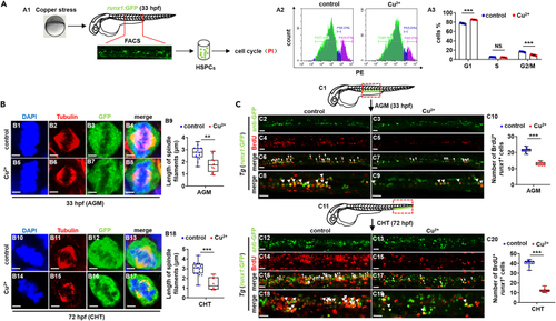
Cu overload induces HSPC proliferation impairment (A) Cell cycle of HSPCs in Cu-stressed embryos at 33 hpf. A1, Schema for the experiments; A2, flow cytometry (FACS) histogram; A3, cell cycle stage calculation. (B) Mitotic malformation of HSPCs in Cu-stressed embryos at 33 hpf (AGM) and 72 hpf (CHT), respectively. B1, B5, B10, B14, DAPI staining; B2, B6, B11, B15, anti-α-tubulin staining; B3, B7, B12, B16, anti-GFP staining; B4, B8, B13, B17, merged. At least 10 mitotic HSPCs in more than 10 embryos were observed for each group. B9, B18, calculation the length of spindles in metaphase runx1GFP+ cells at 33 hpf and 72 hpf, respectively. (C) BrdU cell proliferation assays in embryos at 33 hpf (C2-C9) and 72 hpf (C12-C19), respectively. C1, C11, AGM and CHT domain in embryos, respectively; C10, C20, calculation of the number of proliferative HSPCs in embryos from different groups. C2-C9, C12-C19, lateral view, anterior to the left, and dorsal to the up. Data are mean ± SD. t-test, ∗p < 0.05, ∗∗p < 0.01, ∗∗∗p < 0.001. NS, not significant. Scale bars, 2 μm (B1-B8, B10-B17) and 20 μm (C2-C9, C12-C19). |