
Fig. 2
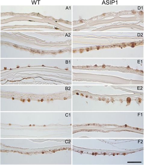
Fig. 2. Presence of lectins in skin. Histological sections showing glucidic residues bound to glycoconjugates in wild type (WT) and ASIP1-overexpressing transgenic zebrafish. The following horseradish peroxidase (HRP) conjugated lectins were used: Canavalia ensiformes/ConA (mannose and/or glucose), Triticum vulgaris/WGA (N-acetyl-d-glucosamine, Ulexeuropeus/UEA-I (l-Fucose), Sambucusnigra/SNA (NeuNAc/sialic acid/NANA) and Glycine max/SBA (α-N-acetyl-d-galactosamine). A) WT/SBA, B) WT/WGA, C) WT/SNA, D) ASIP1/SBA, E) ASIP1/WGA and F) ASIP1/SNA. (1) and (2) indicate dorsal and ventral respectively. Scale bar is 100 μm. |