
Fig. 5
- ID
- ZDB-FIG-230131-16
- Publication
- Ortas et al., 2023 - Label-free imaging of red blood cells and oxygenation with color third-order sum-frequency generation microscopy
- Other Figures
- All Figure Page
- Back to All Figure Page
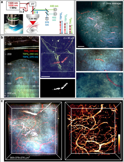
Deep-tissue TSFG imaging in live adult zebrafish brain.
a TSFG microscopy setup scheme for deep tissue imaging with 1 MHz excitation and signal epi-detection. b (left) XZ reslice calculated from a color TSFG z-series spanning >600 μm of imaging depth through the skin, skull and telencephalon of an adult fish. (middle) Sub-skin image showing different spectral signatures for RBCs (in red) and myelinated axons. Segmented RBCs are shown below the image. (Right) Representative images recorded at several depths in the telencephalon, illustrating the specific TSFG contrast of RBCs and vessels. c 3D rendering of the imaged volume in the telencephalon and of the segmented blood vessels. Scale bar: 50 μm. See also Movies M7–10 |