
FIGURE
Figure 4
- ID
- ZDB-FIG-230124-153
- Publication
- Grossi et al., 2022 - Lasp1 Expression Is Implicated in Embryonic Development of Zebrafish
- Other Figures
- All Figure Page
- Back to All Figure Page
Figure 4
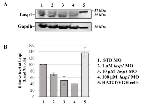
lasp1 MO morphants. (A) Representative Western blotting for Lasp1 protein level in 48 hpf embryos injected with STD MO or lasp1 MO at different doses (1, 10 and 100 µM). Human hepatocellular carcinoma cells (HA22T/VGH) were used as positive controls for LASP1 expression. Gapdh was used as an internal control. The images are representative of two independent experiments. (B) Relative quantifications of Lasp1 expression levels. The histograms represent the mean IOD (integrated optical density) values in percentages; the bars represent SDs from two independent experiments. |
Expression Data
Expression Detail
Antibody Labeling
Phenotype Data
Phenotype Detail
Acknowledgments
This image is the copyrighted work of the attributed author or publisher, and
ZFIN has permission only to display this image to its users.
Additional permissions should be obtained from the applicable author or publisher of the image.
Full text @ Genes (Basel)