
Fig. 4
- ID
- ZDB-FIG-221222-11
- Publication
- Chen et al., 2021 - Acute brain vascular regeneration occurs via lymphatic transdifferentiation
- Other Figures
- All Figure Page
- Back to All Figure Page
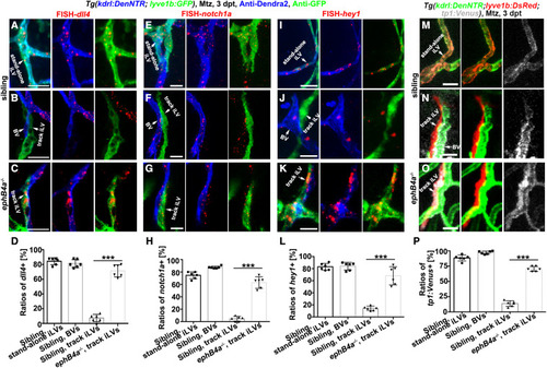
Figure 4. Defective EphB4a leads to derepression of Notch in the track iLVs (A–L) Triple labeling of anti-Dendra2, anti-GFP, and FISH-dll4 (A–C)/notch1a (E–G)/hey1 (I–K). In the siblings, dll4/notch1a/hey1 were activated in the Dendra2+GFP+ stand-alone iLVs (A, E, and I) and Dendra2+GFP− nascent BVs (B, F, and J), but not in the Dendra2−GFP+ track iLVs (B, F, and J). By contrast, in the ephB4a mutant, the Dendra2−GFP+ track iLVs at 3 dpt also exhibited dll4, notch1a, and hey1 expressions (C, G, and K). The statistics show the dll4/notch1a/hey1 expression in the vessels of sibling and ephB4a mutant (D, H, and L) (n = 6 larvae; two-tailed unpaired t test; ∗∗∗, p < 0.0001). (M–P) The Notch functional reporter tp1:Venus was activated in the Dendra2+DsRed+ stand-alone iLVs (M) and Dendra2+DsRed− nascent BVs (N), but not in the Dendra2−DsRed+ track iLVs (N). By contrast, in the ephB4a mutant, the Dendra2−DsRed+ track iLVs at 3 dpt also exhibited Venus expression (O). The statistics show the tp1:Venus expression in the vessels of siblings and ephB4a mutants (P) (n = 6 larvae; two-tailed unpaired t test; ∗∗∗, p < 0.0001). Scale bar, 20 μm. Data are represented as mean ± SD. See also Figures S6 and S7. |
Reprinted from Developmental Cell, 56(22), Chen, J., Li, X., Ni, R., Chen, Q., Yang, Q., He, J., Luo, L., Acute brain vascular regeneration occurs via lymphatic transdifferentiation, 3115-3127.e6, Copyright (2021) with permission from Elsevier. Full text @ Dev. Cell