
Fig. 4
- ID
- ZDB-IMAGE-221222-11
- Publication
- Chen et al., 2021 - Acute brain vascular regeneration occurs via lymphatic transdifferentiation
- All Figures
- Figures for Chen et al., 2021
|
Fig. 4
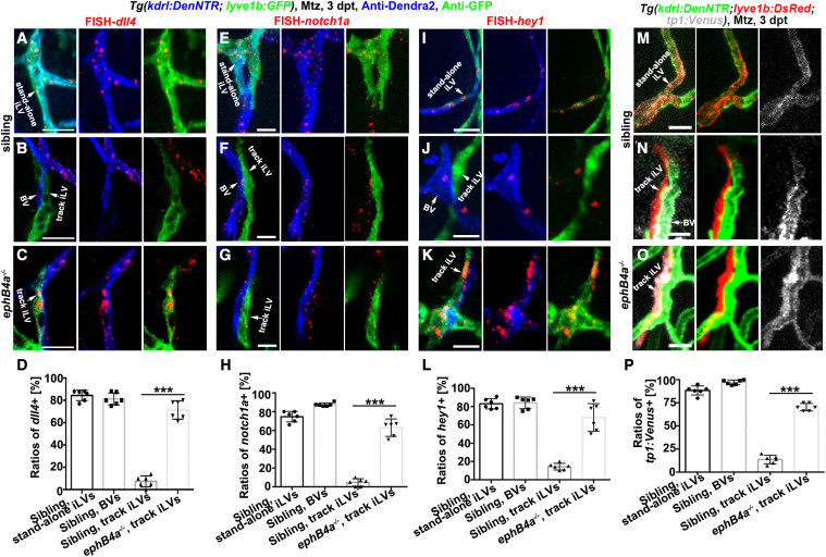
Figure 4. Defective EphB4a leads to derepression of Notch in the track iLVs (A–L) Triple labeling of anti-Dendra2, anti-GFP, and FISH-dll4 (A–C)/notch1a (E–G)/hey1 (I–K). In the siblings, dll4/notch1a/hey1 were activated in the Dendra2+GFP+ stand-alone iLVs (A, E, and I) and Dendra2+GFP− nascent BVs (B, F, and J), but not in the Dendra2−GFP+ track iLVs (B, F, and J). By contrast, in the ephB4a mutant, the Dendra2−GFP+ track iLVs at 3 dpt also exhibited dll4, notch1a, and hey1 expressions (C, G, and K). The statistics show the dll4/notch1a/hey1 expression in the vessels of sibling and ephB4a mutant (D, H, and L) (n = 6 larvae; two-tailed unpaired t test; ∗∗∗, p < 0.0001). (M–P) The Notch functional reporter tp1:Venus was activated in the Dendra2+DsRed+ stand-alone iLVs (M) and Dendra2+DsRed− nascent BVs (N), but not in the Dendra2−DsRed+ track iLVs (N). By contrast, in the ephB4a mutant, the Dendra2−DsRed+ track iLVs at 3 dpt also exhibited Venus expression (O). The statistics show the tp1:Venus expression in the vessels of siblings and ephB4a mutants (P) (n = 6 larvae; two-tailed unpaired t test; ∗∗∗, p < 0.0001). Scale bar, 20 μm. Data are represented as mean ± SD. See also Figures S6 and S7.
Reprinted from Developmental Cell, 56(22), Chen, J., Li, X., Ni, R., Chen, Q., Yang, Q., He, J., Luo, L., Acute brain vascular regeneration occurs via lymphatic transdifferentiation, 3115-3127.e6, Copyright (2021) with permission from Elsevier. Full text @ Dev. Cell