
Fig. 7
- ID
- ZDB-FIG-221222-14
- Publication
- Chen et al., 2021 - Acute brain vascular regeneration occurs via lymphatic transdifferentiation
- Other Figures
- All Figure Page
- Back to All Figure Page
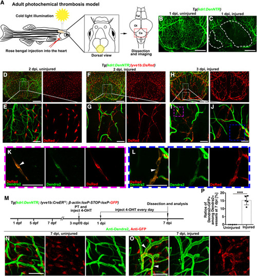
Figure 7. The iLV-to-BV conversion is conserved in the adult photochemical thrombosis model (A) Illustrations of the cold light-induced thrombosis in the zebrafish brain. The yellow circle and red frame indicate the illuminated and imaged cerebellum areas, respectively. Tel, telencephalon; Ot, optic tectum; Ce, Cerebellum. (B and C) In contrast to the uninjured control (B), damage to the cerebellar blood vasculature was observed at 1-day post illumination (C). The dashed lines mark the injured areas (n = 10 adult brains). (D–G) At 2 dpi, in contrast to the interspersed distribution of BLECs on the surface of uninjured brain (D and E), ingrowth of BLECs into the injured area to form iLVs was observed (F and G). The framed areas in (D) and (F) are enlarged in (E) and (G), respectively. n = 10 adult brains. (H–L) At 3 dpi, a portion of lyve1b+ iLVs in the injured cerebellum co-expressed the BEC-specific Dendra2 (K, L, arrowheads). From (H) to (I) and (J), then to (K) and (L), sequential enlargements of the framed areas are shown. n = 10 adult brains. (M–P) Transgenic lines, time points of photochemical thrombosis (PT) and 4-OHT administration (M). In contrast to the uninjured adult brain (N, injected rose bengal without cold light illumination, then injected 4-OHT from 0 to 7 dpi every day), a portion of BVs were double positive for anti-GFP and anti-Dendra2 in the injured brain at 7 dpi (O, arrowheads). The statistics show the ratios of Dendra2+GFP+ vessels among all the Dendra2+ blood vessels at 7 dpi (P) (n = 5 regions from 5 different adult brains; two-tailed unpaired t test; ∗∗∗, p < 0.0001). Scale bars in (B), (C), (D), (F), and (H): 200 μm; Scale bars in (E), (G), (I–L), (N), and (O): 50 μm. Data are represented as mean ± SD. |
Reprinted from Developmental Cell, 56(22), Chen, J., Li, X., Ni, R., Chen, Q., Yang, Q., He, J., Luo, L., Acute brain vascular regeneration occurs via lymphatic transdifferentiation, 3115-3127.e6, Copyright (2021) with permission from Elsevier. Full text @ Dev. Cell