
Fig. 7
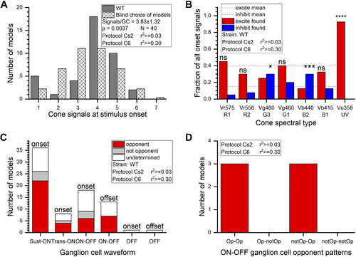
Cone input patterns in onset responses. A: the number of cone signals found for ON responses in “best” ganglion cell (GC) models varied from 1 to 6, with a peaked distribution centered at 3.8 (dark bars). The distribution differed from blind choices among the 127 models [hatched bars, χ2(6,40) = 19.27, P ≤ 0.01]. Models are numbered from 1 to 127, with the group of single-cone models on left and the single-membered 7-cone group on right. The “blind” choice distribution is proportional to the number of models in each group. B: overall patterns of excitation and inhibition found at stimulus onset differed among spectral types of cone. The fraction of excitatory (red) or inhibitory (blue) signals found for the 40 “best” onset models is illustrated for each cone type. For all cones, the fraction of modeled excitation was 0.40 (dashed red line). The fraction of modeled inhibition was 0.15 (dashed blue line). The significance of the ways individual cone types differed from these overall means is indicated by asterisks (χ2 tests, see text). Vu358 (UV) cone signals were significantly different, being 93% excitatory, whereas Vg480 (G3) green cones and Vb440 (B2) blue cones favored inhibition. C: in sustained-ON (Sust-ON), transient-ON (Trans-ON), and ON-OFF GC types, modeled opponency was the most common pattern, and nonopponency was rare. “Undetermined” models are those that did not meet the criterion r2 values. D: for ON-OFF GCs where opponency could be determined for both ON and OFF signals, it was common that both phases were color opponent, but the type of opponency, or nonopponency, differed. WT, wild type. |