
Fig. 1
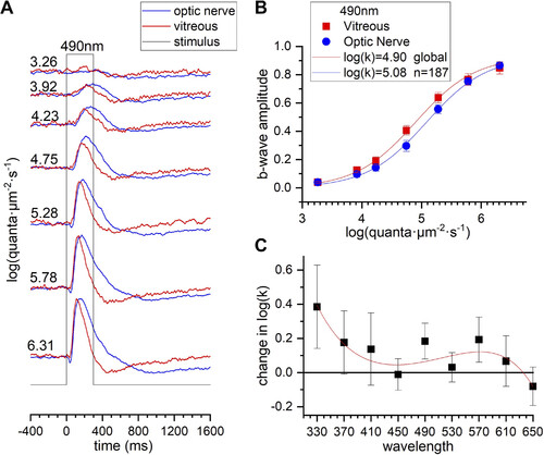
Absorption of spectral stimuli by larval sclera, choroid, and pigment epithelium. Electroretinogram (ERG) b wave sensitivity is used to estimate stimulus attenuation with transscleral stimuli. A: recordings within the optic nerve, using transscleral stimulation (blue), and within the vitreous, using corneal stimulation (red), are normalized to the maximal amplitude at each recording configuration and superimposed. The quantal irradiance, given on left of each response trace in log(quanta·µm−2·s−1), is calibrated at the eye surface. B: maximum-amplitude-normalized data sets at 490 nm for 8 vitreal and 21 optic nerve b wave data sets are combined and globally fit by Hill functions. The semisaturation irradiance k is fit separately, but the saturation amplitude Vm and exponent n are constrained to be the same. Overall, vitreal b waves were 0.18 log units more sensitive than optic nerve b waves at 490 nm. C: the change in log(k) across stimulus wavelengths is fit by a 4th-order polynomial. In B and C error bars are SEs. |