
Fig. 6
- ID
- ZDB-FIG-221030-6
- Publication
- Ferre-Fernández et al., 2022 - CRISPR-Cas9-mediated functional dissection of the foxc1 genomic region in zebrafish identifies critical conserved cis-regulatory elements
- Other Figures
- All Figure Page
- Back to All Figure Page
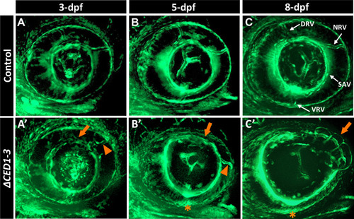
foxc1a∆CED1−3 mutant embryos display defects in the developing superficial choroidal vasculature. A–C’ Three-dimensional maximum intensity projection images of the ocular vasculature in live control (A–C) or foxc1a∆CED1−3 homozygous (A’–C’) embryos carrying fli1a:EGFP transgene at 3-, 5- and 8-dpf. Mutant embryos show abnormal development of the dorsal and nasal radial vessels (orange arrowheads in A’–C’), enlarged and deformed superficial annular vessel (orange arrows) and a highly disorganized vasculogenesis in the ventral part of the eye with no visible ventral radial vessel at 5- and 8-dpf (orange asterisks). DRV (dorsal radial), NRV (nasal radial), SAV (superficial annular), and VRV (ventral radial) blood vessels are indicated |