
FIGURE
Fig. 5
- ID
- ZDB-FIG-221023-17
- Publication
- Liu et al., 2022 - Lrpap1 deficiency leads to myopia through TGF-β-induced apoptosis in zebrafish
- Other Figures
- All Figure Page
- Back to All Figure Page
Fig. 5
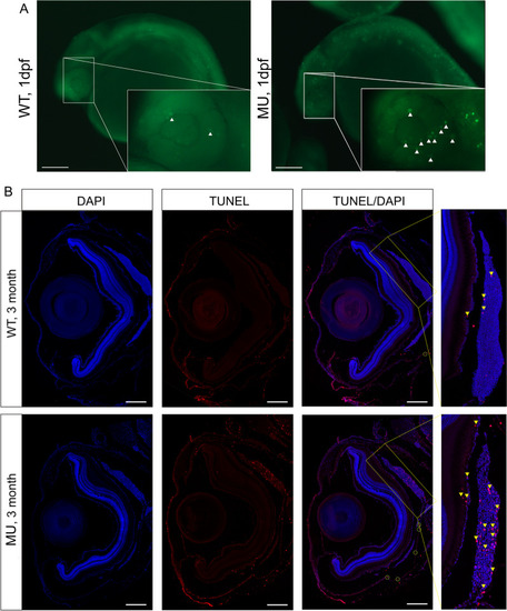
Knockout of lrpap1 in zebrafish leads to increased apoptosis in the eye. (A) AO was used to quantify apoptosis in the eyes of 24 hpf embryos: green fluorescence, as highlighted using white triangles. (B) TUNEL staining was also used to investigate ocular apoptosis in adult zebrafish. Apoptosis is highlighted by yellow triangles, particularly in the choroidal tissue (magnified image). The green circles highlight apoptotic cells in the sclera. The scale bars refer to 200 μm. WT, wild-type. MU, lrpap1 homozygous mutant. dpf, days post-fertilization |
Expression Data
Expression Detail
Antibody Labeling
Phenotype Data
| Fish: | |
|---|---|
| Observed In: | |
| Stage: | Prim-5 |
Phenotype Detail
Acknowledgments
This image is the copyrighted work of the attributed author or publisher, and
ZFIN has permission only to display this image to its users.
Additional permissions should be obtained from the applicable author or publisher of the image.
Full text @ Cell Commun. Signal.